Tips and Tricks in ECG Diagnosis
Case 1 - Nonbacterial Thrombotic Endocarditis
A 57-year-old man presented with confusion, cough, fever, and unintentional weight loss. Before this presentation, he had malaise and anorexia developed. Also, a nonproductive cough developed, along with fevers to a maximum temperature of 37.6°C. On clinical examination, the temperature was 38.3°C, the heart rate 111 beats per minute, the blood pressure 135/73 mm Hg, the respiratory rate 24 breaths per minute, and the oxygen saturation 94% while he was breathing ambient air. Laboratory test results were notable for a troponin I level of 13.4 ng per milliliter (reference range, <0.04), a lactate level of 2.4 mmol per liter (22 mg per deciliter [reference range, 0.4 to 2.0 mmol per liter; 6 to 18 mg per deciliter]), and a white-cell count of 21,800 per cubic millimeter (reference range, 4500 to 10,500). An electrocardiogram (ECG) showed sinus rhythm at 99 beats per minute, with 1‑mm concave ST‑segment elevations and PR‑segment depressions in leads 2, 3, and aVF; subtle PR‑segment elevation in lead aVR; and submillimeter upsloping ST‑segment depressionsin lateral and anterior leads. The findings could indicate pericarditic or ischemic changes.
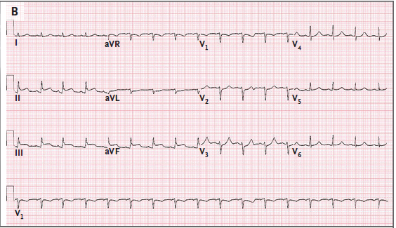
Q. What is the most probable diagnosis?
Discussion
Nonbacterial thrombotic endocarditis is considered to be an uncommon condition, and it is most often found post mortem, with rates in autopsy series ranging from 0.9 to 1.6%. This immune-mediated phenomenon is characterized by the presence of sterile vegetations, composed of bland fibrin–platelet thrombi that form on valvular leaflets. It typically occurs in patients with advanced cancer (known as marantic endocarditis) and in those with systemic lupus erythematosus (known as Libman–Sacks endocarditis). However, it may also complicate other chronic diseases, such as tuberculosis, uremia, and the acquired immunodeficiency syndrome. The most common initial manifestation of nonbacterial thrombotic endocarditis is an embolic event that results in neurologic symptoms Patients with nonbacterial thrombotic endocarditis tend to have evidence of multiple, widely distributed brain infarcts on diffusion-weighted MRI, whereas single lesions or focal infarcts are more characteristic of infective endocarditis. The vegetations that result from nonbacterial thrombotic endocarditis are typically friable and have a high propensity for embolization because of the minimal inflammatory reaction at the site of attachment. They are classically found along valvular coaptation lines (i.e., where the valve edges meet) and are not associated with destruction of valvular tissue. The vegetations are typically small (<1 cm in diameter), broad-based, and irregular in shape, and they are frequently located on the mitral and aortic valves; cases of nonbacterial thrombotic endocarditis involving valves on the right side of the heart are rare.
N Engl J Med. 2018 Oct 25;379(17):1658-1669.
Case 2 - Pericardial Effusion
A 16-year-old girl presented for syncope and altered mental status. She began to complain of chest pain, diaphoresis, and nausea. She vomited, became clammy, and fell to the ground unresponsive. She remained groggy, tachycardic, and anuric. On physical examination, she is sleepy but responsive, with a Glasgow Coma Scale score of 15, delayed capillary refill, 2+ pulses, and hepatomegaly to 4 cm below the costal margin. She denies pain, fevers, night sweats, unintended weight loss, exercise intolerance, or peripheral edema. Her vital signs showed, temperature, 97.5ºF (36.4ºC); heart rate, 120 beats/min; respiratory rate, 23 breaths/min; blood pressure, 95/44 mm Hg; and oxygen saturation, 98% on room air. Initial laboratory results show metabolic acidosis with venous pH 7.29; carbon dioxide level, 38 mEq/L (38 mmol/L); white blood cell count, 28,800/mL (28.8 x 10⁹/L); lactate level, 40.5 mg/dL (4.5 mmol/L); blood glucose level, 187 mg/dL (10.4 mmol/L); troponin I level, 0.04 ng/mL (0.04 mg/L); brain-type natriuretic peptide level, 42 pg/mL (42 ng/L); and cortisol level, 49 mg/dL (1,352 nmol/L). electrocardiogram shows PR depression showing pericardial inflammation/pericarditis, and also shows right axis deviation concerning for right heart strain.
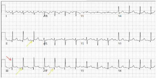
Q. Most probable diagnosis?
Discussion
In a pediatric population, vasovagal syncope and orthostatic hypotension are more common than cardiac causes of syncope. Cardiac (electrical and structural) causes of syncope are rare but can be life-threatening. A pericardial effusion refers to the accumulation of fluid in the pericardial sac surrounding the heart. The pericardial sac is composed of the thin visceral pericardium which consists of a single layer of cells adherent to the cardiac epicardium and the thicker and fibrous parietal pericardium composed of collagen and elastin which is adherent to the lungs, diaphragm, sternum, great vessels, and other mediastinal structures surrounding the heart. In a healthy individual, the pericardial sac contains between 15 mL and 50 mL of serous fluid. Pericardial effusion is possible across all ages and populations. The predominant etiology of the effusion varies by demographic characteristics such as age, geography, and comorbidities. The ECG findings vary from normal to non-specific ST segment changes for small effusions. For large effusions or tamponade, the ECG may demonstrate electrical alternans which is a specific but not a sensitive finding. If the pericardial effusion is due to pericarditis, then the ECG may show PR depressions or diffuse ST elevations.
Pediatr Rev. 2019 Jan;40(1):37-39.
Pericardial Effusion. [Updated 2018 Dec 19]. In: StatPearls [Internet].
Case 3 - Lyme Carditis
A 28-year-old presented with a non-prodromal syncope episode and a 4-day history of chest pain, shortness of breath, and malaise. His vital signs and physical examination were within normal limits. His laboratory results showed elevated inflammatory markers with a CRP of 94.5 mg/L and ESR of 44 mm/hr, as well as mildly elevated troponin of 51 ng/L. He had a history of of a tick bite 1–2 monthsprior. Lyme disease serology was taken. An echocardiogram (ECG) showed a junctional rhythm at 70 bpm and it also showed mild global hypokinesis mostly affecting the inferior and infero-septal walls.
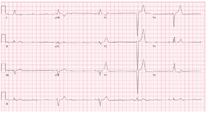
Q. What is the most Probable diagnosis?
Discussion
Lyme disease is a bacterial infection caused by Borrelia burgdorferi, transmitted through certain Ixodes tick species. Affected patients can present with erythema migrans (EM), arthralgias, neurologic symptoms, and systemic symptoms such as malaise and fatigue. When the bacteria invade the heart, it can lead to Lyme carditis. Lyme carditis is a rare complication of the B. burgdorferi infection affecting approximately 10% of reported cases. Symptoms include chest pain, shortness of breath, palpitations, dizziness, and syncope as about 90% of patients with Lyme carditis will present with high-degree AV block. Diagnosis often proves to be challenging as many patients do not remember a previous exposure to tick bites and do not have EM. The use of the Suspicious Index in Lyme Carditis (SILC) score may help identifying Lyme carditis. Most patients with Lyme carditis present with high-degree AV block, it is important to recognize this reversible cause of heart block to prevent unnecessary permanent pacemaker implantation. Pathologic features of Lyme carditis in humans have been described in small numbers and it showed infiltration of the endocardium, myocardium, and pericardium with lymphocytes, plasma cells, and macrophages. Perivascular inflammation has been prominent in most cases. Typically advanced heart block and positive Lyme serology bring most patients to proper diagnosis, other ECG changes may be witnessed, including a wide spectrum of AV block. Because of the rarity of this diagnosis, and given the frequent absence of other concurrent clinical manifestations of early Lyme disease, consideration of Lyme carditis demands a high level of suspicion when patients in endemic areas come to attention with cardiovascular symptoms and evidence of higher-order heart block.
Ann Noninvasive Electrocardiol. 2019 May;24(3):e12599.
Infect Dis Clin North Am. 2015 Jun;29(2):255-68.
Case 4 - Bundle branch reentrant Ventricular Tachycardia (BBRVT)
A 52-year-old man was presented with a history of ischemic cardiomyopathy (ICM). He had a left ventricular (LV) ejection fraction of 15–20% also had a single chamber primary prevention ICD placed. He was presented with frequent ICD shocks. On clinical examination, a unique, fast (200–300 beats/min), monomorphic wide complex tachycardia (WCT) associated with syncope, hemodynamic compromise, and cardiac arrest was recorded. He underwent multi-vessel coronary artery bypass grafting (CABG). After a year of ICD implantation, the patient received seven appropriate ICD shocks for episodes of monomorphic Ventricular tachycardia (VT). He experienced a total of 40 episodes of tachyarrhythmias labeled by his device as either SVT or VT for which he has received 28 ICD shocks. The electrocardiogram (ECG) revealed sinus rhythm, left superior axis, and left bundle branch block (LBBB) of 134-ms duration.
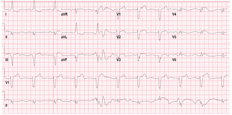
Q. What is the most Probable diagnosis?
Discussion
Bundle branch reentrant ventricular tachycardia (BBRVT) is characterized by a unique, fast (200–300 beats/min), monomorphic wide complex tachycardia (WCT) associated with syncope, hemodynamic compromise, and cardiac arrest. BBRVT is more common in NICM patients, is also seen in ICM patients with scar in the interventricular septumIt is often challenging to diagnose usually requiring a His bundle recording and specific pacing maneuvers. The overall incidence has been reported to be between 3.5 to 6% of VTs in different series, and up to 20% among patients with non-ischemic cardiomyopathy (NICM) undergoing electrophysiologic studies (EPS) and ablation. There are three categories of BBRVT, Type A and C is the classic counterclockwise and clockwise BBRVT circuits, respectively. Type B is interfascicular reentry within the LBB fascicles themselves, most commonly seen in patients with anterior wall infarction and left anterior or posterior hemifascicular block. Patients with BBR commonly have prolongation of the PR interval, QRS duration, and HV interval. BBR can also occur in patients with normal His-Purkinje conduction during sinus rhythm but impaired at faster rates.
J Interv Card Electrophysiol. 2018 Aug;52(3):385-393.