Tips and Tricks in ECG Diagnosis
Case 1 - Uhl's anomaly
A 17-year-old boy presented with facial puffiness and swelling in the lower limbs for 6 months and one episode of syncope 15 days earlier. There was no significant family history of cardiomyopathy, congestive heart failure, or sudden cardiac death. His blood pressure was 106/70mm Hg, heart rate 102 beats min-1,oxygen saturation on room air was 100%. A 12-lead electrocardiogram showed sinus rhythm with incomplete right bundle branch block. The right atrium was dilated and hypertrophied. There was no fat signal in the right ventricular wall, in contrast to that seen in arrhythmogenic ventricular dysplasia. The tricuspid valve leaflets were in their normal anatomical position. Cardiac magnetic resonance imaging showed an extremely dilated thin-walled right ventricle and absence of trabeculations. The septal and left ventricular myocardium was preserved.
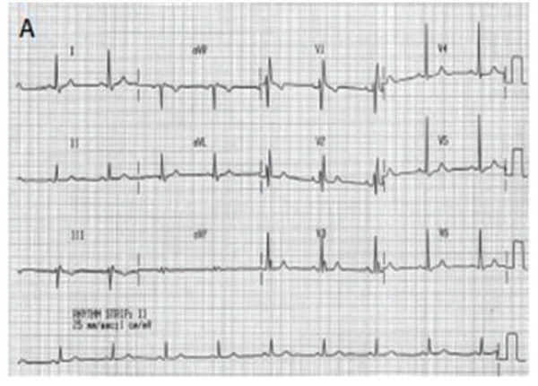
Q. What is the most probable diagnosis?
Discussion
Uhl’s anomaly is a rare entity characterized by absent myocardium in the right ventricle with apposition of the epicardium and endocardium. It most commonly presents as congestive heart failure and arrhythmias. Uhl’s anomaly is one of the differential diagnoses in right ventricular dilatation, which include Ebstein’s anomaly, partial anomalous venous return, absence of the pericardium, pulmonary atresia, and right ventricular arrhythmogenic dysplasia. Morphological features of Uhl’s anomaly include complete absence of the myocardium in the right ventricular parietal wall, which is composed of the endocardium and epicardium, with no interposition of adipose tissue between these layers. The male sex predominates in Uhl’s anomaly,with an incidence of 56%.Previously, the diagnosis of Uhl’s anomaly was usually obtained at autopsy, but with the development of echocardiography and magnetic resonance imaging, early diagnosis is possible.
Asian Cardiovasc Thorac Ann.2018 Sep;26(7):563-565.
Case 2 - Cardiac amyloidosis
A 77-year-old woman admitted to the hospital with anorexia, dyspnea on exertion and recurrent syncope for 1 month. Physically, she had edema in both legs and macroglossia, but showed no jugular venous distention. There were neither respiratory wheezes nor cardiac murmurs heard. On admission, her blood pressure was 88/58mmHg, heart rate 75/min. The blood cell count revealed hypocytosis, hemoglobin of 79 g/L, red blood cell count of 2.5×10¹²/L, white blood cell count of 3.0×10⁹/L, platelet count of 66×109/L, and elevated erythrocyte sedimentation 57mm/h. Urine analysis was positive for protein, the excretion of 24 hours urinary protein is 2570mg/L. The 12-lead ECG revealed low-voltage QRS complexes in the limb leads, normal voltages and poorly progressive R wave in the precordial leads.
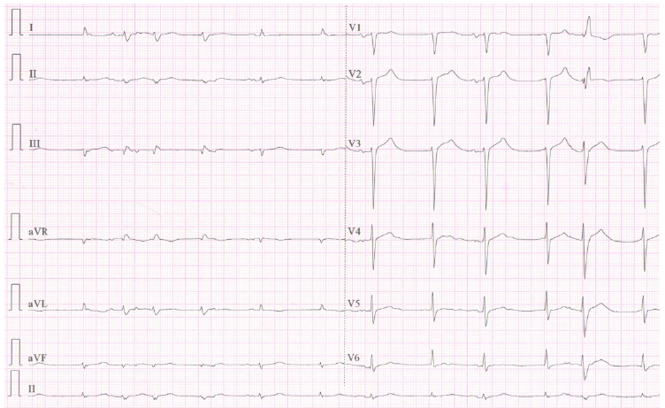
Q. Most probable diagnosis?
Discussion
The amyloidosis is a systematic disease with the heterogeneity of cardiac, renal or skin involvement. Cardiac amyloidosis is an infiltrative Cardiomyopathy, presenting as restrictive Cardiomyopathy or hypertrophic Cardiomyopathy. The contradiction between echocardiographic hypertrophy and low voltages in the limb leads in ECG is the obvious feature of cardiac amyloid. Systemic amyloidosis generally results from widespread deposition of insoluble fibrillar proteins, like free light chains with the heterogeneity of cardiac, renal or skin involvement. Amyloidosis is rarely associated with multiple myeloma. Cardiac amyloidosis is an uncommon cause of heart failure. In this patient, Congo red staining of the abdominal fat biopsy was positive, which was the gold standard for diagnosis of amyloidosis. The electrocardiogram demonstrates low QRS voltages in the limb leads with a poorly progressive R wave and normal voltages in the precordial leads is suggestive of an infiltrative process. Biatrial enlargement is the feature of restrictive Cardiomyopathy and resulted from diastolic dysfunction. The contradiction between echocardiographic hypertrophy and low voltages in the limb leads in ECG is the significant feature of cardiac amyloid and diagnostic clues.
QJM. 2019 Feb 1;112(2):131-132.
Case 3 - Congenital inferior sinus venosus defect
A 21-year-old man presented to the emergency department with dizziness and worsening dyspnea on exertion. Patient has no past medical history. Physical examination was significant for grade III/VI systolic murmur over the left 2nd intercostal space. Electrocardiography showed sinus rhythm and right ventricular hypertrophy (RVH). Transthoracic echocardiography (TTE) revealed preserved left ventricular ejection fraction, RVH, and dilated right chambers. Pulmonary valve leaflets appeared thickened with restricted systolic excursion correlating with pulmonary valve Stenosis. Peak pressure gradient across the pulmonic valve was 39 mm Hg. Right ventricular systolic pressure was estimated to be 69 mm Hg based on a measured tricuspid regurgitation velocity of 3.9 m/s.
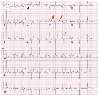
Q. What is the most Probable diagnosis?
Discussion
Congenital inferior sinus venosus defect (SVD) is a rare type of atrial Septal defect (ASD) that is located in the inferior portion of the atrial septum. This defect is commonly misdiagnosed as secundum ASD with inferior extension. Using the strict anatomic and echocardiographic definition of SVD is crucial in distinguishing inferior SVD from other subtypes of ASD, especially secundum ASD. While secundum ASD is amenable to transcatheter device closure, inferior SVD requires surgical repair. The absence of the posterior rim in the short-axis view can help to differentiate inferior SVD from a secundum ASD. Although the use of multiple modalities such as transesophageal echocardiography, computed tomography, and MRI could help to clarify some of the anatomical distinctions between different defects, their use is not necessary to make the diagnosis. Inferior SVD can alter the hemodynamics with consequent increase in pulmonary pressure leading to early onset dyspnea on exertion, fatigue, and recurrent respiratory tract infections. Pulmonary valve Stenosis should be considered in the setting of RVH in young patients before estimating pulmonary artery systolic pressure; failure of which may result in an erroneous diagnosis of pulmonary hypertension.
Echocardiography. 2018 Aug;35(8):1227-1229.
Case 4 - Allergic myocardial infarction(Kounis syndrome)
A 57-year-old woman presented to Emergency department(ED) with urticarial rash on her face, precordial pain, and palpitations 5 minutes after the first subcutaneous injection of 10 IU recombinant human insulin. The patient had never experienced a severe allergic reactionin the past. The medical history of the patient revealed that she had a history of hypertension, hyperlipidemia, and type 2 diabetes for the past 10 years and started to insulin therapy on the day of the ED presentation. Vital signs were as follows: temperature 37.2 °C, heart rate 102 beats per minute,blood pressure 150/70 mm Hg, respiratory rate 28 breaths per minute, and pulse oximetry 96% on room air. Her electrocardiogram showed ST segment elevations in leads V1-V5. Transthoracic echocardiography, revealed anterior wall hypokinesia with a left ventricular ejection fraction of 54%.
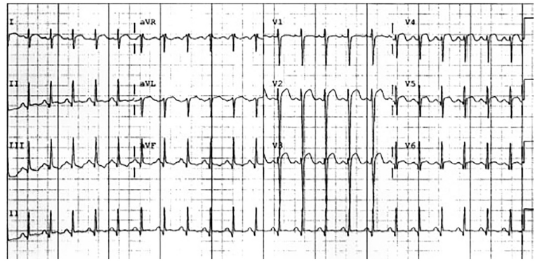
Q. What is the most Probable diagnosis?
Discussion
Allergic angina and allergic myocardial infarction, currently known as Kounis syndrome, is defined as acute coronary syndrome caused by an allergic reaction or a strong immune reaction to a drug or other substance. Kounis syndrome is caused by inflammatory mediators such as histamine, neutral proteases, arachidonic acid products, and a variety of cytokines and chemokines released, during mast cell degranulation. Kounis syndrome has been linked with several diseases (bronchial asthma, urticaria, food allergy), environmental exposures (viper, wasp or bee venoms) and a variety of drugs used widely in daily clinical practice, such as antibiotics, analgesics, antineoplastics, contrast media, corticosteroids, intravenous anesthetics, non-steroidal anti-inflammatory drugs and others. But Kounis Syndrome secondary to insulin has not been reported before. The diagnosis of Kounis Syndrome is based on clinical symptoms and signs as well as on laboratory, electrocardiographic, echocardiographic, and angiographic evidence. All patients admitted to the emergency department with chest pain and ST elevation on electrocardiography should be asked about allergic insults.
Heart Lung. 2018 Jul -Aug;47(4):360-362.