Tips and Tricks in ECG Diagnosis
Case 1
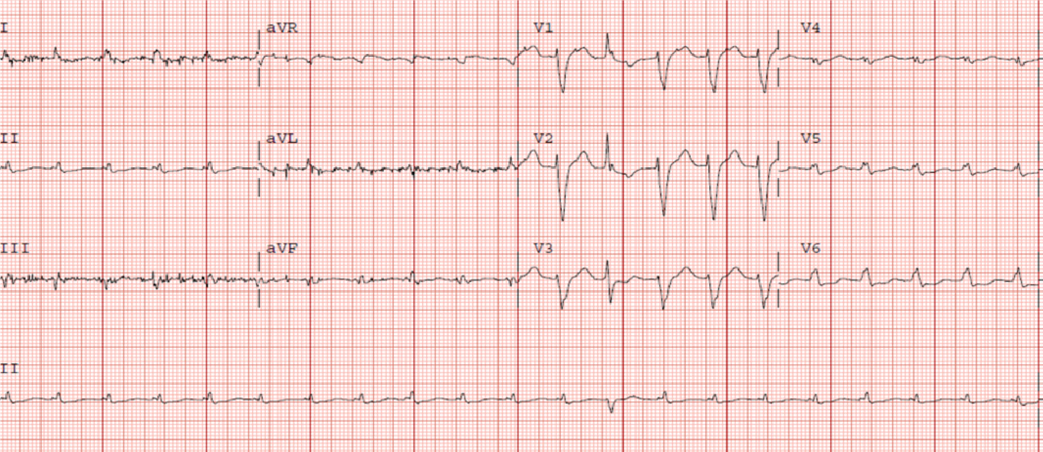
A 73 years old female presented to the emergency department with sudden onset shortness of breath with no associated chest pain. She was emergently intubated due to impending respiratory failure. Her medical history was notable for congestive heart failure, atrial fibrillation, primary hyperparathyroidism, carpal tunnel syndrome and spinal stenosis. On initial physical examination, she had a blood pressure of 80/54 mmHg with pulse rate of 75 beats per minute (bpm). Her jugular venous pressure was not elevated. She had clear lungs and normal heart sounds with no murmurs or gallops on auscultation. Her lower extremities were cool to touch with mild pitting edema bilaterally. Laboratory testing was significant for N-terminal pro b-type natriuretic peptide (NTProBNP) elevation at 12914 pg/mL (Normal < 125 pg/mL). Electrocardiogram (ECG) showed left bundle branch block (LBBB) and low voltage in the limb leads while the chest radiograph demonstrated bilateral patchy opacities consistent with pulmonary edema. Transthoracic ECHO revealed a borderline dilated left ventricle (LV) with severe left ventricular hypertrophy and reduced LVEF of 30%-35%. There was discordance between the degree of LV thickness on ECHO and QRS voltage on ECG. An assessment of cardiogenic shock was made and inotrope therapy with intravenous dobutamine was initiated. She progressed well with addition of isosorbide dinitrate/hydralazine (40-75 mg) twice a day to her medication regimen prior to discharge home. Neurohormonal blocking agents for her systolic dysfunction were continued with titration of her diuretic depending on her volume status.
Q. What is the most probable diagnosis?
Discussion
Transthyretin amyloidosis (ATTR) occurs due to the misfolding of the liver derived precursor protein transthyretin (TTR, previously called prealbumin). Wild type ATTR was previously described to be exclusive to older white males. However, more cases in females are being identified as in this patient suggesting this condition may under-diagnosed in this population. The diffuse deposition of amyloid fibrils in the cardiac interstitial space produces significant thickening of both ventricles with associated stiffness which results in impaired diastolic relaxation and the characteristic restrictive physiology. Diagnosing cardiac amyloidosis requires a high index of suspicion and a combination of laboratory tests, ECG, cardiac imaging and histopathology is required. The first clue which may prompt suspicion for cardiac amyloidosis is the ECG. The archetypal finding is low QRS voltages due to the electrically silent amyloid fibrils which are not detected by the ECG. This is inconsistent with the degree of ventricular thickening seen on ECHO. The absence of low QRS voltage does not preclude a diagnosis of cardiac amyloidosis with only 30%-70% of patients meeting low voltage criteria on ECG. A pseudoinfarct pattern with Q waves in the early precordial leads is another common ECG finding, identified in about 40%-50% of cardiac amyloidosis patients. Other ECG findings frequently encountered include atrial arrhythmias such as atrial fibrillation, various degrees of atrioventricular block, interventricular conduction delays and bundle branch blocks.
World J Clin Cases. 2019 Mar 26;7(6):742-752.
Case 2
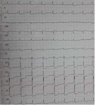
A 68-year-old man who had a history of aortic insufficiency and ascending aortic dilatation for 5 years was admitted to emergency department with exertional dyspnea and coughing. He had no history of hypertension or diabetes mellitus. The physical examination revealed a blood pressure of 100/50 mmHg and a heart rate of 80 beats/min. Blood pressure values were similar between the right and left arms. Femoral, tibialis posterior, and dorsalis pedis pulses were palpable and similar between the right and left limbs. A 3/6 diastolic decrescendo murmur was heard along the left sternal border. Electrocardiography (ECG) showed normal sinus rhythm without any ST segment or T-wave changes. Increased cardiothoracic index and prominent aortic arch were observed on chest X-ray examination. While awaiting the results of laboratory tests, patient described severe chest pain. A repeated ECG showed ST segment elevation in aVR lead and diffuse ST segment depressions in all other derivations. His blood pressure was 90/50 mmHg and heart rate was 100 beats/min. Transthoracic echocardiography revealed severe aortic regurgitation, dilated ascending aorta with a diameter of 5.27 cm, and intimal dissection flap that was prolapsing into the left ventricle during diastole. Left ventricular ejection fraction was found within the normal limits. Immediate surgery was suggested for the patient just before a sudden cardiopulmonary arrest. Cardiopulmonary resuscitation was performed; however, patient passed away.
Q. Most probable diagnosis?
Discussion
Acute aortic dissection (AD) is a life-threatening condition requiring immediate assessment and therapy. The symptoms of A) may be highly variable and may mimic other much common conditions. Thus, a high index of suspicion should be maintaned, especially when the risk factors for AD are present or signs and symptoms suggest this possibility. However, sometimes AD may be asymptomatic or progression may be subclinical. Although most patients with type-B AD are hypertensive, several patients with type-A AD are normotensive or hypotensive on admission. The present patient was normotensive on admission; however, his hemodynamic status was deteriorated in a short time period. The ECG changes in AD are usually non-specific; however, approximately 1%–2% of the patients have acute ST segment elevation. Studies have reported that the most common ECG changes in patients with acute AD were ST segment depression or T-wave changes. Although ST segment elevation in aVR lead is not a common ECG sign in patients with acute AD, diffuse ST segment depression with ST segment elevation in aVR lead was appeared just after the beginning of severe chest pain in the present patient. In such patients, ST segment elevation in aVR lead may reflect the occlusion of left main coronary artery orifice. In patients with a history of aortic insufficiency and/or enlargement of ascending aorta when admitted to the emergency departments with sudden chest pain, acute AD should be considered in the differential diagnosis.
Interv Med & App Sci. 2019;11 (1):68–70.
Case 3
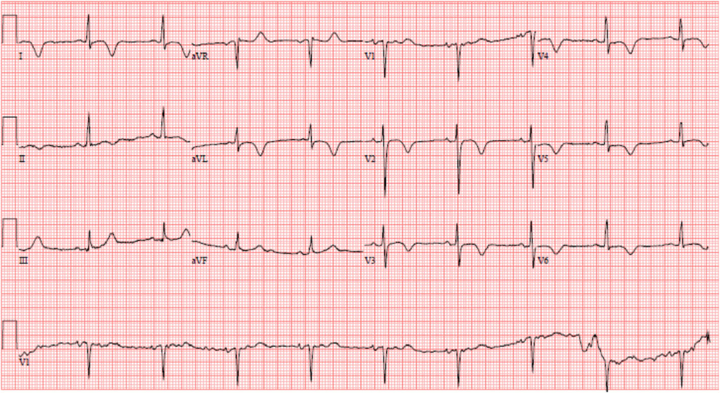
A 54 year-old male with a history of orthotropic heart transplant 17 years ago for non-ischemic cardiomyopathy presented to the hospital after experiencing intermittent palpitations with associated lightheadedness of one hour duration. He denied any chest pain or shortness of breath, orthopnea or paroxysmal nocturnal dyspnea. At baseline, he described activities consistent with New York Heart Association Class 2. Additional medical history was notable for benign prostatic hyperplasia and hyperlipidemia. He denied tobacco or illicit drug use and alcohol consumption. He had a blood pressure of 184/89 mmHg on arrival but otherwise had stable vital signs and physical examination. Cardiac biomarkers revealed a CK of 59 U/L and a troponin of 0.11NG/ML (normal < 0.04NG/ML). B type natriuretic peptide was 371 PG/ML (normal 0-100PG/ML). An electrocardiogram revealed a normal sinus rhythm with T wave inversions in the anterior and lateral leads (leads V2 to V6) which were unchanged compared to prior EC. He had no evidence of arrhythmias on cardiac monitoring. Transthoracic echocardiogram revealed left ventricular ejection fraction of 55–65%. His cardiac biomarkers trended down to normal and he had no evidence of arrhythmias on continuous cardiac monitoring so he was discharged from the hospital with close follow up with his heart transplant team.
Q. What is the most Probable diagnosis?
Discussion
Apical Hypertrophic Cardiomyopathy (ApHCM) is a relatively rare variant of hypertrophic cardiomyopathy associated with thickening of the apical segment of the left ventricle leading to a ‘spade-shaped’ small cavity. Initially described in Japan, it makes up approximately 15– 25% of all hypertrophic cardiomyopathies and is more commonly seen in men. The hallmarks include giant T wave inversions in the precordial leads on electrocardiogram and the aforementioned spade like appearance of the left ventricular cavity. Clinically, the manifestations can vary from apical aneurysms, atrial tachycardia or atrial fibrillation, ventricular arrhythmias and sudden cardiac death. About 54% of patients are asymptomatic. Nevertheless, one third of patients experience serious cardiovascular complications, such as myocardial infarction and arrhythmias. The finding of apical hypertrophic cardiomyopathy in this patient likely does not suggest an increase in mortality or morbidity due to its historically benign course. However, recent studies have shown increased morbidity and mortality in this subset of patients. Factors like advanced age, hypertension, diabetes, baseline atrial fibrillation, left atrial volume index and apical aneurysm on cardiac magnetic resonance are predictors of poor prognosis. The etiology of ApHCM in this patient is thought to be idiopathic.
BMC Cardiovasc Disord. 2019 Apr 3;19(1):81.
Case 4
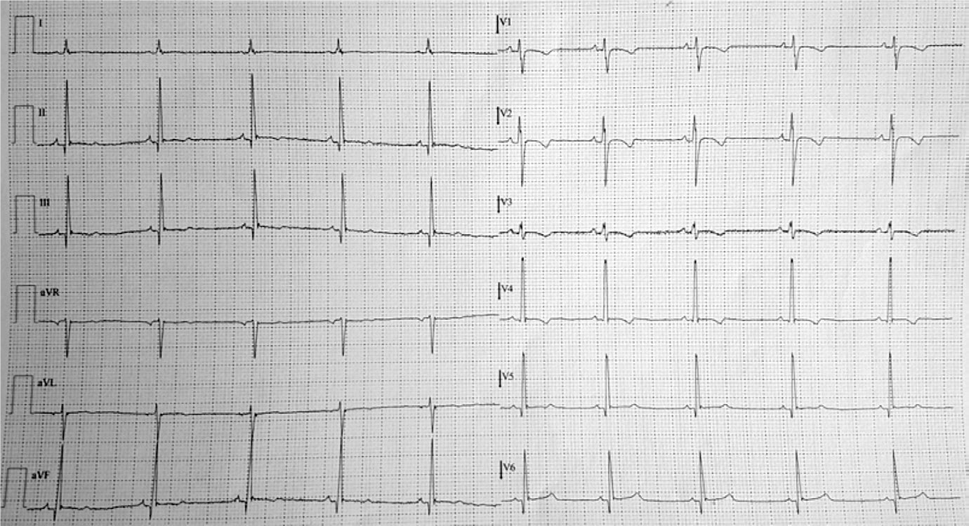
An 18-year-old male arrived at the ED after experiencing an unprecedented sudden and unexpected syncope with retrograde amnesia and without loss of sphinterial function during a basketball match. Once admitted, the patient underwent computer tomography scan of the brain, electroencephalogram and a complete neurologic examination to rule out ischemic events or epilepsy. These tests came back with negative results. Medical physical examination showed normal systolic and diastolic blood pressure heart rate, body temperature of 36°C, oxygen saturation on air at 99% and no signs or symptoms of heart failure. A 12 leads electrocardiogram (ECG) showed regular sinus rhythm at 70 bpm with T-wave inversion in the anterior leads (from V1 to V4), mild T-wave abnormalities in peripheral leads and incomplete right bundle branch block. No previous ECG of the patients was available. A complete echocardiographic evaluation was performed and showed normal left ventricular (LV) size and ejection fraction (EF: 56%); normal thickness of the interventricular septum and posterior wall but with thinning of the apical segment of the lateral wall that appeared hypokinetic. Systolic function was globally reduced with fractional area shortening of 38% and global strain - 10%; evident trabeculation of the free wall with a “stack of plates” shape in mid-basal segment Given this clinical scenario, the patient underwent subcutaneous defibrillator (ICD) implantation. Sotalol 80 mg daily was started accordingly.
Q. What is the most Probable diagnosis?
Discussion
Arrhythmogenic right ventricular cardiomyopathy (ARVC) is a rare cardiomyopathy characterized by a progressive replacement of the myocytes by various degrees of fibro-adipose tissue involving particularly the areas between the anterior part of the pulmonary infundibulum, the apex, and the infero-posterior wall of the RV. Although up to 76% of the ARVC hearts studied at post-mortem disclosed a LV involvement, the biventricular diseases is less common in vivo. These abnormalities can lead to sudden cardiac death and/or heart failure. Similarly to this case, T-wave inversions in the right precordial leads are present in up to 87% of adult patients with ARVC due to RV dilatation. However, they can be found in other contexts, such as in some young athletes, in ischemic cardiomyopathy, or during acute pulmonary embolism. Incomplete or complete right bundle branch block is also described and manifested in this patient. Epsilon wave (electrical potentials of small amplitude that occur at the end of the QRS complex and at the beginning of the ST segment in the right precordial leads) represents areas of delayed activation frequently localized in the RV as a consequence of fibrous and/or fibro-fatty replacement of myocardium and it is considered a major criterion for the diagnosis of ARVC. Although the clinical course of the disease is known to be largely unpredictable, with such extensive morphological abnormalities malignant arrhythmias are often early symptomatic.
Monaldi Archives for Chest Disease 2019; 89:1009