Tips and Tricks in ECG Diagnosis
Case 1
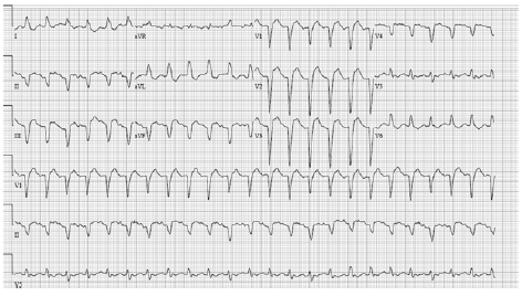
A 37-year old woman was admitted to the hospital for treatment of symptomatic VT that was frequent and refractory to untiarrhvthmic agents. ECG recorded during the VT showed a left bundle branch block pattern in the precordial leads and an inferior axis. This pattern fulfilled the criteria of VT originating from the aortic sinus cusp. ECG recorded during sinus rhythm showed a normal QRS morphology and ST-T segment. Extensive investigation including echocardiography, left and right ventriculography, and coronary angiography revealed no structural heart disease. During rapid pacing from the midseptal RV during the VT. Classic entrainment criteria, including constant fusion and pro-gressive fusion, were fulfilled, suggesting that the mechanism of VT was reentry with an excitable gap. Double potentials preceding the QRS complex by 58 ms were recorded from the posterior RV outflow tract (RVOT). The first component of the double potentials was captured orthodromically by pacing from the midseptal RV at a pacing rate of 225 beats/min becau.se the morphology of the first component during pacing was the same as that during the spontaneous VI, and the time interval between the last electrogram activated by the pacing stimulus to the initial electrogram of the spontaneous VT after pacing was equal to the pacing cycle length. In contrast, the morphology of the second component of the double potentials during pacing was different from that during the spontaneous VT. When detailed analysis of the conduction time was performed during VT entrainment. a slow conduction zone was detected between the midseptal RV and posterior RV, because the posterior RV was captured by pacing from the midseptal RV with a low conduction time.
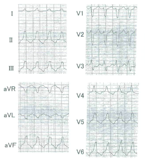
Q. What is the most probable diagnosis?
Case 2
A 56-year-old woman was evaluated for a 2- year history of recurrent syncope preceded by nausea, chest pain, and palpitations. Her history showed neither cardiovascular events nor internal disease, aside from hypercholesterolemia. Both her parents had suffered myocardial infarction at the age of 50. All findings derived from physical examination, laboratory tests, chest X ray, echocardiography, Doppler, and cardiac catheterization (including right- and left-sided pressure measurements, coronary arteriography, and biplane cineangiography of both ventricles) were normal. The 12-lead ECG during sinus rhythm showed abnormalities specific for the Brugada syndrome with a RBBB morphology, left-axis deviation, QRS width of 160 ms, and QTc of 440 ms. Leads V1 and V2 showed 0.2-mV ST-segment elevation. Shortly after admission the patient had a spell of syncope with transiently reduced consciousness. The ECG showed a monomorphic VT; rate 25D/min. The VT had a left bundle branch block (LBBB) morphology and superior axis. The VT was preceded by recurrent ventricular extrasystoles morphologically identical to that of the VT. Because the VT persisted, and the patient remained in a reasonably good hemodynamic condition (blood pressure 110/70 mmHg), conversion was attempted by intravenous (IV) procainamide. After 200 mg of procainamide, tbe VT terminated and sinus rhythm was restored. On the EGG, the QRS duration was then 180 ms, QTc 475 ms, and Vi and V2 showed more prominent (0.5-mV) ST elevation. Interestingly, two shortly coupled ventricular extrasystoles occurred with LBBB configuration and concordant QRS morphology in Vi-Ve, proving a ventricular origin. The EGG abnormalities diminished during the washout of procainam.ide. Two days later, the patient had several episodes with cardiac arrest. Two-channel EGG recordings now showed rapid polymorphic VT deteriorating into VF that required electrical cardioversion. Initiation of polymorphic VT was preceded by sinus rhythm with single and double, shortly coupled extrasystoles. In the following 24 hours, the patient had several episodes of nonsustained polymorphic VT.
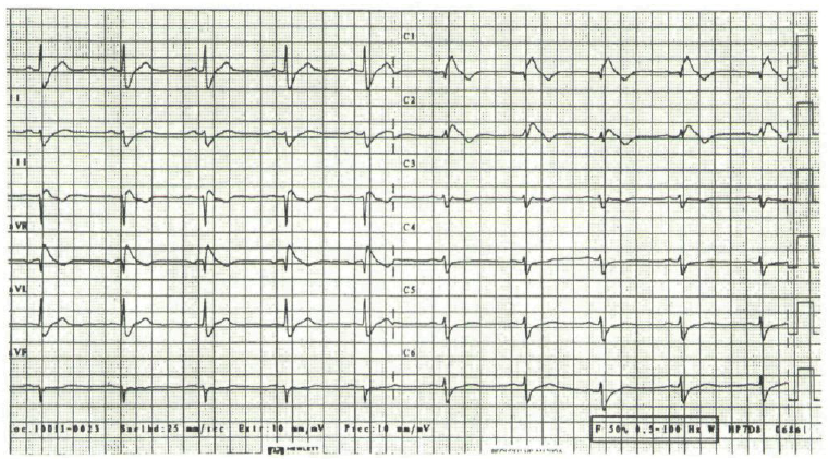
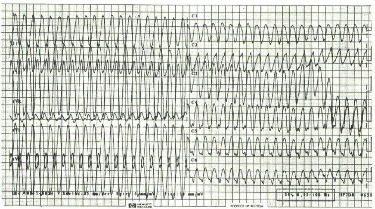
Q. What is the most probable diagnosis?
Case 3
An 18-year-old woman was referred to an Intensive Care Unit (ICU) after being resuscitated from cardiac arrest. She had a history of congenital disorder, characterized by facial dysmorphism, deafness, and vesicoureteral. There was no history of cardiomyopathy or cardiac disease. She suddenly developed cardiac arrest at home. Cardiopulmonary resuscitation was initiated by paramedics 5 minutes after syncope. The use of a semiautomatic defibrillator revealed ventricular fibrillation. Electric cardioversion (200 J monophasic) was recommended, restoring sinus rhythm. Mechanical ventilation was initiated, and the patient was transferred to our ICU. On admission, blood pressure was 100/57 mmHg, heart rate 100 beats/min, and no cardiac murmur or abnormal heart sound were detected. The 12-lead ECG revealed normal sinus rhythm andWolff-Parkinson- White syndrome, due to a left accessory pathway. The QT interval was normal. The chest x-ray was normal. Transthoracic and transesophageal echocardiography revealed a normal size left ventricle (LV) with a normal left ventricular systolic ejection fraction. Nevertheless, echocardiography disclosed several abnormalities. There was thin compacted myocardium on the epicardial side of left ventricle and thicker noncompacted myocardium on the endocardial side. Numerous trabeculae were found located in the apical segment of the LV. The ratio of noncompacted/ compacted myocardium measured at endsystole was 2.6. Recesses filled with blood from the LV cavity were visualized by color Doppler imaging. These findings are characteristic discriminatory features, leading to diagnosis of isolated left ventricular noncompaction, in the absence of any major associated cardiac abnormalities.
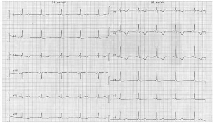
Q. What is the most Probable diagnosis?
Case 4
A 72-year-old previously healthy woman was hospitalized for high-grade fevers (2-week history) unresponsive to acetaminophen and for profound weakness, myalgia, headaches, and poor oral intake. She reported no recent contact with sick people, no recent history of travel, no history of blood transfusion, and no smoking, alcohol, or recreational drug use. Her medical history was significant for hypothyroidism treated with levothyroxine. Examination at presentation was unexceptional apart from high-grade fevers up to 104 °F. Her initial laboratory studies showed a white blood cell count of 7.6 ×10³/μL (normal range, 4–11 ×103/μL) with a lymphocyte count of 66% (normal range, 20%– 53%). Her C-reactive protein level was 11 mg/dL (normal, ≤0.49 mg/dL). Her liver enzymes on admission were elevated: alkaline phosphatase, 201 U/L (normal range, 20–140 U/L); aspartate aminotransferase, 171 U/L (normal range, 5–40 U/L); and alanine aminotransferase, 162 U/L (normal range, 5–40 U/L). Her bilirubin lev- els were normal. Chest radiography showed bibasilar opacities. Urinalysis results were normal. A computed tomogram (CT) of the chest, abdomen, and pelvis, and ultrasonograms of the right upper quadrant were also normal. Blood, urine, and respiratory cultures were negative. Because of the persistent high-grade fevers, there was extensive investigation of possible causes—including an influenza A/B enzyme immunoassay and tests for Epstein-Barr virus, CMV DNA polymerase chain re-action (PCR), parvovirus B19 antibodies, anaplasma phagocytophilia antibodies, hepatitis B virus, hepatitis C virus, HIV, and rheumatic diseases. All the tests were negative, except for a CMV immunoglobulin M (IgM) titer that was strongly positive at 1.75 (index value, <0.9) with a normal CMV IgG titer of 0.6 (index value, <0.8), which suggested an acute CMV infection. A CMV DNA quantitative PCR assay revealed 13,100 viral copies/mL (>1,000 copies/mL is high). After diagnosis, the patient was started on oral valganciclovir. Within 48 hours after initiation of treatment, despite decreased viral counts, the patient developed sudden-onset shortness of breath and was noted to be febrile (temperature, 102 °F), tachycardic (heart rate, 142 beats/min and regular), tachypneic (respiratory rate, 32 breaths/min), hypotensive (blood pressure, 72/54 mmHg), and hypoxemic (SaO2, 68% on room air). Cardiovascular examination revealed distended jugular veins and an S3 gallop. Lung auscultation revealed bibasilar rales. A chest radiograph showed prominent interstitial markings and hilar congestion that suggested pulmonary edema and small bilateral pulmonary effusions. A CT angiogram confirmed pulmonary interstitial edema with moderate-sized bilateral pleural effusions and showed significant cardiomegaly, when compared with the CT scan performed on admission. An electrocardiogram (ECG) showed sinus tachycardia and previously existing left bundle branch block. There was also mild elevation in cardiac troponin T to 0.36 ng/mL (normal, <0.03 ng/mL), with normal creatine kinase levels. The patient’s brain natriuretic peptide level was elevated at 1,537 pg/mL (normal, <100 pg/mL). Echocardiography showed severe global hypokinesis with a left ventricular ejection fraction (LVEF) of 0.25, and tissue Doppler imaging was consistent with diastolic dysfunction. The diagnosis of CMV-induced fulminant myocarditis was made on the basis of the acute clinical presentation and serologic evidence of infection.
Q. What is the most Probable diagnosis?