Tips and Tricks in ECG Diagnosis
A 50-year-old female with retrosternal pain and dyspnea
A 50-year-old, obese female, was admitted after four episodes of syncope accompanied by retrosternal pain and dyspnea. There was no history of diabetes or arterial hypertension, but she reported being an ex-smoker (tobacco load of 60 packs per year) and undergoing radiotherapy for uterus cancer 10 years before. The report of her admission was as follows: tachycardia (152 bpm); blood pressure of 133/74 mmHg; and O2 saturation of 92% at room air. Her heart rhythm was irregular, with neither accessory heart sounds nor murmurs. The respiratory sounds were reduced in the lower third of both hemithoraces. The ECG on admission showed atrial fibrillation with ventricular response of 150 bpm, S1Q3 pattern, incomplete right bundle-branch block (RBBB), ST-segment elevation in the leads aVR and V1, and ST-segment depression in the leads D1, D2, aVF, and V2-V6. The markers of myocardial necrosis were positive and the troponin I peak was 4.42. The patient underwent coronary angiography, which showed no coronary artery obstruction. A transthoracic echocardiogram revealed normal left and right ventricular (RV) function, while venous Doppler echography of the lower limb evidenced recent thrombosis of the popliteal and small saphenous veins.
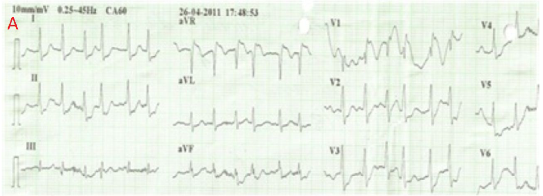
Q. What is the diagnosis?
Discussion
The classical ECG changes of pulmonary thromboembolism (PTE) are as follows: T wave inversion on the right precordial leads; simultaneous T wave inversion and ST-segment elevation on the anteroseptal and inferior leads; S1Q3T3 pattern; complete or incomplete RBBB; and sinus tachycardia. The presence of atrial arrhythmias, RBBB, and ST-segment elevation or depression is associated with RV failure and poor prognosis. The ECG changes of PTE are attributed to changes in the cardiac hemodynamics. Volume overload increases the parietal tension and the RV muscle work. The ventricular septum bulging into the left ventricle reduces cardiac output, decreasing coronary flow, which, associated with hypoxemia and the increased heart work, contributes to myocardial ischemia, justifying the elevation of myocardial necrosis markers. According to her Wells score (4.5 points), the patient here reported had an intermediate probability of PTE: 3 points for the clinical symptoms of deep venous thrombosis (asymmetric swelling with tenderness on palpation) and 1.5 points for heart rate greater than 100 bpm. Adding the ECG changes found increased the probability of PTE. However, the ST-segment elevation in the leads aVR and V1 could also indicate severe damage to the left main coronary artery and/or proximal lesion of the anterior descending artery. Thus, the presence of the ST-segment elevation in the leads aVR and V1 could mean both massive PTE and severe coronary artery lesion.
Source: Arq Bras Cardiol 2012; 99(3): e131-e133
A 42-year-old woman with swollen and tender right feet
A 42 year old woman presented with acute, painful, right leg swelling beginning five hours earlier that morning after waking. During the examination she noted sudden abdominal pain, became short of breath, and had a syncopal episode for 20 seconds. The right leg was swollen and tender but well perfused and the abdomen was painful with guarding and rebound. The initial 12 lead ECG showed a sinus tachycardia of 120 beats/min with ST elevations in V1 to V3, S wave and ST depression in D1, ST depression in D2, ST elevation in aVR, ST depression in aVL, Q wave and ST elevation in V1, ST elevation in V2 and V3, and ST depression in V5 and V6. Cardiac ultrasound done in the emergency department showed a normally contracting left ventricle, a distended right ventricle with free wall hypokinesia, and displacement of the interventricular septum towards the left ventricle.
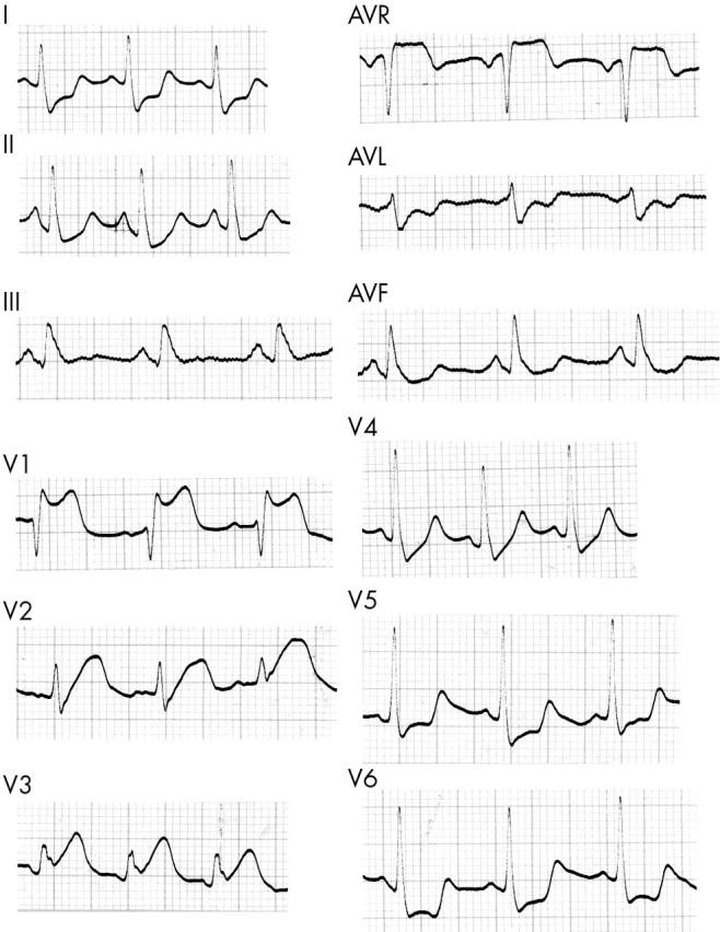
Q. What is the diagnosis?
Discussion
Pulmonary embolism (PE) is most often a complication of venous thrombosis, usually of the lower extremities and the pelvis. Massive PE is life threatening if left untreated, with rapid progression and deterioration. ECG abnormalities can be seen in 70–80% of patients, varying from the typical S1Q3T3 pattern to no specific changes. Very rarely ECG mimics anteroseptal infraction. The ECG is often abnormal in PE but the findings are insensitive and non-specific. ECG findings are usually non-specific and almost 33% of patients have a normal ECG. The most common findings are sinus tachycardia and non-specific ST segment and T wave changes. Changes that strongly suggest PE indicate strain on the right side of the heart; these changes include T wave inversion in precordial leads V1 through V4, transient right bundle branch block (RBBB), new right or left deviation of the QRS axis, sudden onset of atrial fibrillation or other atrial arrhythmias, and ECG signs of right ventricular hypertrophy or right atrial enlargement. The S1Q3T3 pattern also suggests PE. This pattern of right sided heart strain is usually accompanied by T wave inversion in the precordial leads. In the present case the ST elevation was confined to the anteroseptal wall, the axis was normal, there were S waves in leads I and aVL > 1.5 mm, the transaction zone was in V3, and there was ST depression in V6.
Source: Heart. 2004; 90(7): e41
A 13-year-old girl with an episode of generalized tonic-clonic seizures.
A 13-year-old girl presented to the emergency department with an episode of generalized tonic clonic seizures. Postictal, patient was drowsy but hemodynamically stable. Postictal monitoring followed by a 12-lead electrocardiogram (ECG) showed fast atrial fibrillation (AF) at 200 beats per minute (bpm) with intermittent wide QRS regular tachycardia suggestive of monomorphic ventricular tachycardia (VT) at 280 bpm. Immediately following this, her rhythm changed to wide QRS irregular tachycardia suggestive of polymorphic VT at 220 bpm without hemodynamic compromise. At times, the ventricular rate exceeded 300 to 350 bpm. Subsequent electrocardiogram in sinus rhythm showed short PR of 130 milliseconds, wide QRS of 140 milliseconds, and deltawaves in the QRS. There was dominant R in the right precordial leads and negative delta waves in leads II, III, and aVF (pseudoinfarct pattern), indicating a left posteroseptal accessory pathway. General as well as cardiovascular examination was unremarkable. Her computed tomography scan of head and results of blood investigations including electrolytes did not show abnormalities.
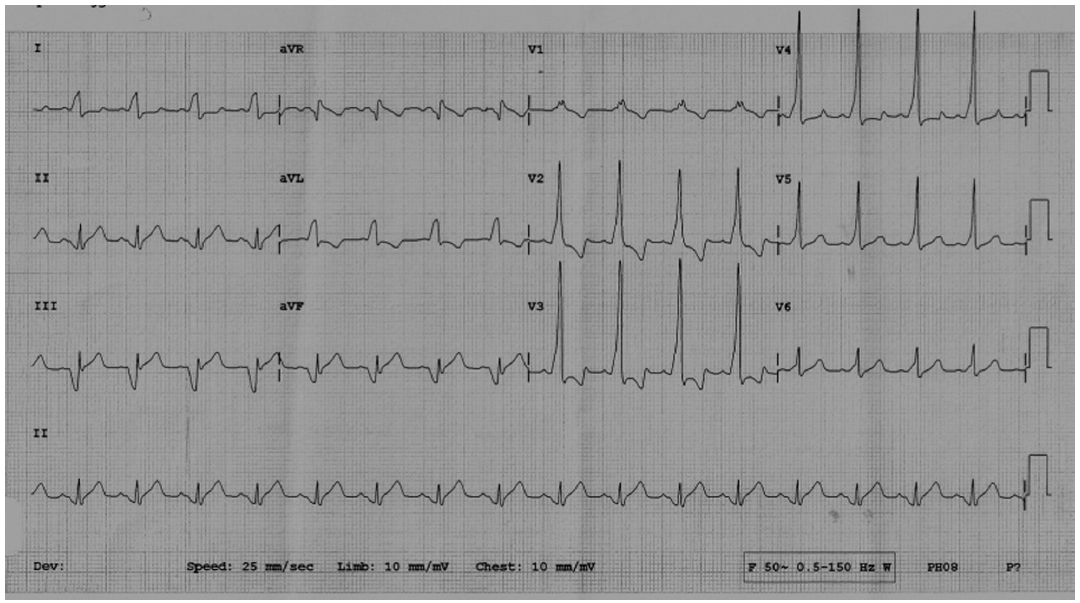
Q. What is the diagnosis?
Discussion
This patient presented with multiple narrow QRS and wide QRS tachyarrhythmias. In classic Wolff-Parkinson-White (WPW) syndrome, during sinus rhythm there is fast antegrade conduction over the accessory pathway compared with slower atrioventricular (AV) node conduction resulting in early depolarization of the ventricles manifesting short PR, wide QRS, and delta wave on ECG. Wide QRS tachycardia can occur in patients with antidromic AV reciprocating tachycardia (AVRT) or dual pathways (including nodoventricular pathways) or if pre-existing bundle-branch block coexists. In the case presented, initially there was fast AF with intermittent regular wide QRS tachycardia. This regular wide QRS tachycardia was thought to be a VT. Actually, this represented either AVRT or atrial flutter with 1:1 antegrade conduction through the accessory pathway and retrograde conduction through AV node (antidromic conduction). This was followed by wide QRS irregular tachycardia masquerading a polymorphic VT. Again, this represented antidromic conduction of all AF impulses through the accessory pathway leading to wide QRS irregular tachycardia without the QRS twisting around the isoelectric baseline. In this patient at times, the ventricular rate exceeded 300 to 350 bpm, which increases risk of sudden death from ventricular fibrillation. It has been observed that accessory pathway conduction may be intermittent with resultant antidromic AVRT producing wide QRS regular tachycardia or irregular tachycardia in patients with AF. Another interesting aspect in this patient was occurrence of generalized tonic-clonic seizures, which may just be an association with AF or secondary to fast tachyarrhythmias causing Stokes-Adams-Y-type syncope with seizures (aborted VF).
Source: Pediatr Emer Care. 2012; 28(11): 1227-1229
A 65-yr-old man with a laryngeal tumor
A 65-yr-old, 158-cm, 59-kg man with a laryngeal tumor was admitted to hospital for laryngeal microsurgery. He had been diagnosed as having intermittent Wolff-Parkinson-White (WPW) syndrome 2 year previously from a history of palpitations with ventricular pre-excitation on ECG. Disopyramide had been prescribed and he had been well since then. Preoperative 12-lead ECG showed normal sinus rhythm with a delta wave, at a rate of 72 beat/ min. Clinical examination, chest x-ray and routine laboratory investigations were normal. When he was connected to ECG lead II on arrival in the operating room, the ECG showed a normal sinus rhythm with a rate of 82 beat/ min and a narrow QRS complex. General anesthesia was induced and an uneventful operation was performed which lasted for 15 minutes. Three minutes later, the ECG showed conversion of the normal sinus rhythm with a rate of 87 beat/ min to a rate of 80–120 beat/ min. The width of the QRS complex remained normal (Fig 1). Arterial pressure was 151/88 mm Hg. The patient was haemodynamically stable with normal AV conduction. Two minutes after administration of neostigmine, the ECG showed that narrow QRS complex had converted to a wide QRS complex with a rate of 110–180 beat/ min (Fig 2). Systolic arterial pressure decreased to 76 mm Hg.
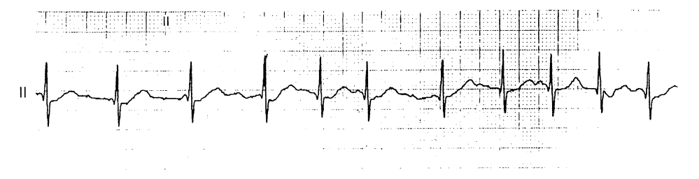
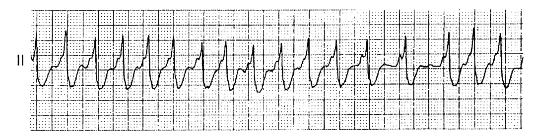
Q. What is the diagnosis?
Discussion
WPW syndrome manifest on the ECG as a short PR interval, a wide QRS complex and a delta wave. One major problem associated with the anesthetic management of patients with WPW syndrome is the risk of tachyarrhythmia as a result of the presence of the accessory pathway. Awareness of the electrophysiological effects of anesthetic and antiarrhythmic drugs on normal AV and accessory pathway conduction is important to avoid tachyarrhythmia in these patients. In this patient, administration of neostigmine converted narrow QRS complexes to wide ventricular complexes and accelerated the ventricular response during atrial fibrillation (AF). The cause of a widening QRS complex can be explained by an inhibitory effect of neostigmine on the normal AV. Therefore, there is a risk that administration of neostigmine during AF in patients with WPW syndrome may lead to rapid ventricular rates that may result in ventricular fibrillation. The use of neostigmine should be avoided during AF associated with WPW syndrome. Recurrent paroxysmal AF has been reported in approximately 32% of patients with WPW syndrome. The accessory pathway properties and atrial vulnerability predispose to AF in patients with WPW syndrome. Increased sympathetic can induce AF by affecting both factors. Failure to treat AF may have allowed progression to wide QRS complex tachycardia after administration of neostigmine. AF should be treated rapidly to restore sinus rhythm before administration of an anti-cholinesterase. In addition, if antagonism of a non-depolarizing neuromuscular blocking drug is indicated during normal sinus rhythm, an anti-cholinesterase drug should be used with caution as autonomic nervous system imbalance during anesthesia in these patients may convert sinus rhythm to AF.
Br J Anaesth 1999; 83(5): 815–18