Tips and Tricks in ECG Diagnosis
Case 1
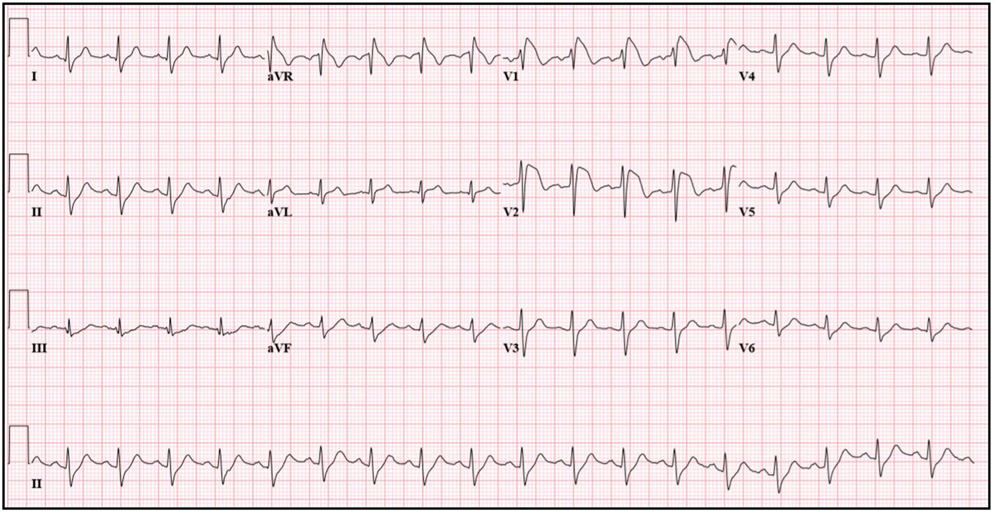
A 47-year-old man with no known past medical history presented to emergency department with chest pain, coughing, and fever up to 104°F for 5 days. His father had died in his 50s of what was assumed to be a myocardial infarction. He did not smoke or use drugs and drank minimal alcohol. With his symptoms not improving, the patient went to a local emergency room. An electrocardiogram showed ST changes that were concerning for an acute ST-elevation myocardial infarction. ST elevation was prominent in leads V1 and V2. He had chest pain, which was more consistent with chest wall pain from recent intractable cough, but given the patient's family history, electrocardiographic changes, and symptoms, he was transferred for a higher level of care and an urgent invasive cardiac workup. Coronary angiography demonstrated no significant coronary narrowing. Echocardiography showed no structural or functional abnormalities. Acetaminophen and ibuprofen were scheduled to lower his ongoing fever. The patient was outside the window for Tamiflu, and his fever gradually subsided with supportive care. With resolution of his fever, his electrocardiogram evolved over the next several days. The patient was counseled on aggressive treatment of fevers in the future and avoidance of potential arrhythmia-inducing drugs. Electrophysiology was consulted, and the patient was deemed to be low risk for sudden death. No implantable defibrillator was indicated.
Q. What is the most probable diagnosis?
Discussion
This case illustrates how undiagnosed Brugada Syndrome (BS) may be unmasked by fever in an adult patient—something that is more commonly seen in the pediatric population. Our patient presented with an electrocardiogram consistent with a type I Brugada pattern in the setting of fever. A study found that fever-induced BS is significantly more likely to present as a type I electrocardiographic pattern than other triggers of Brugada signs and symptoms. This same study found that febrile patients are less likely to have findings of arrhythmia or history of syncope at presentation; rather, the discovery of the Brugada pattern on electrocardiogram is an incidental finding in most of these patients. In general, BS develops in patients with a cardiac channel mutation that causes a transmural voltage gradient between the epicardium and endocardium. Several studies have noted that BS is one of many diagnoses that can be misdiagnosed as ST elevation myocardial infarction (STEMI). The typical electrocardiographic pattern of type I Brugada is a down-sloping ST-segment elevation peaking at 2mm with J wave amplitude in leads V1–V3 and a negative T wave in the same leads; right bundle branch or pseudoright bundle branch block is typically seen in two or more of V1–V3. This should be contrasted with the typical electrocardiographic findings in a patient presenting with STEMI, in which ST segments tend to be convex upward, forming a dome, often exceeding the height of QRS, with widened or inverted T waves, reciprocal ST depression, and Q waves. As this case demonstrates, patients presenting with ST elevation that does not fit the classic pattern associated with STEMI should be evaluated for other causes of electrocardiographic changes.
Proc (Bayl Univ Med Cent). 2018 Nov 2;32(1):107-109.
Case 2
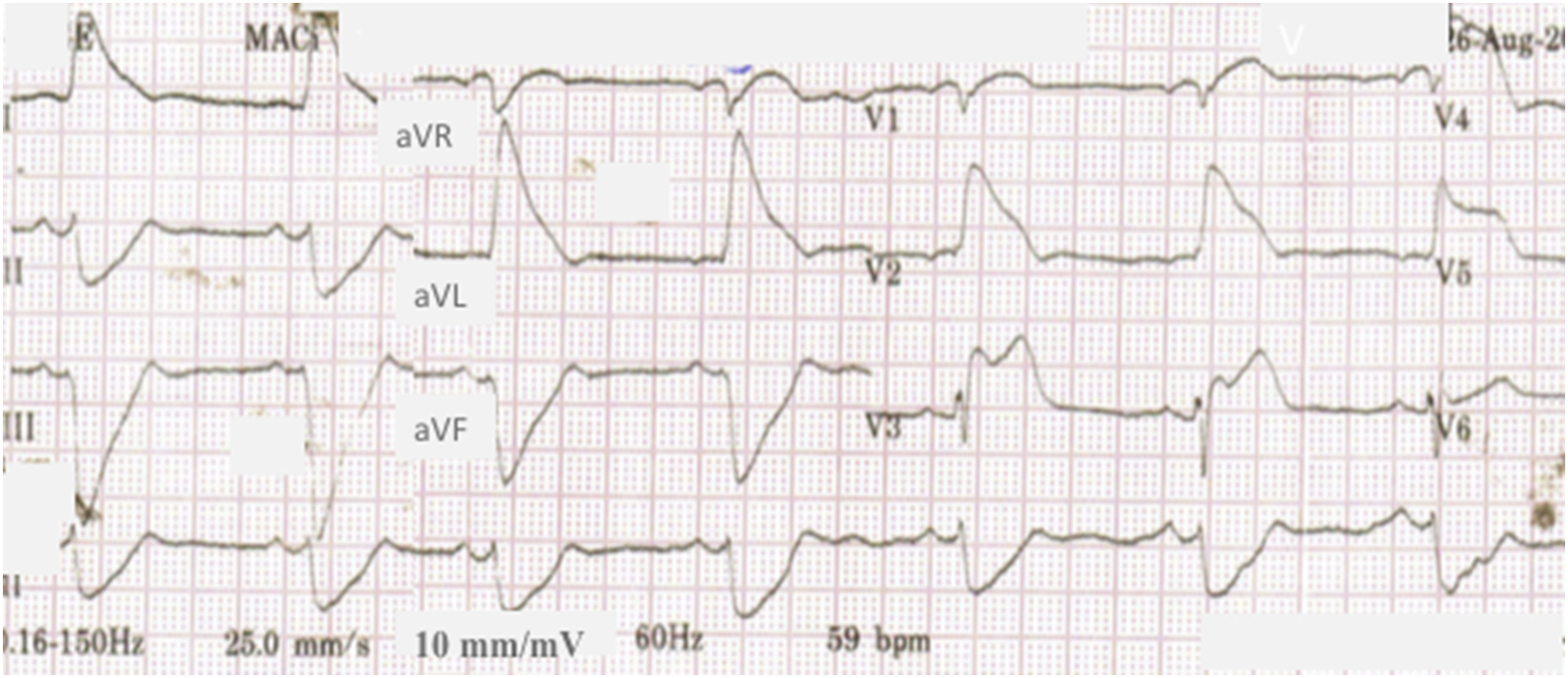
A 54-year-old man presented to the Hospital Emergency Unit with intense chest pain which started 40 minutes before, with irradiation to the upper left arm, associated with nausea and sweating. He had a medical history of diabetes mellitus, dyslipidemia and prior tobacco use. Physical examination showed blood pressure of 90/50 mm Hg, heart rate of 50 bpm, and normal chest auscultation. ECG (Figure 1) showed sinus rhythm with a rate of 56 bpm, T wave inversion in leads II, III, and aVF, and downslopping ST depression with negativepositive biphasic T waves in V3-6. The patient improved and remained asymptomatic for about 4 hours, when he had a new episode of severe chest pain. The ECG then showed sinus rhythm, HR of 59 bpm, and marked ST segment elevation in leads V2 to V6, I and aVL, with evident fusion of QRS complex and T wave. The pain relieved spontaneously in 7 minutes, and resolution of the TW changes occurred 9 minutes after. The patient was referred to the hemodynamics laboratory. Drug-eluting stent implantation was performed uneventfully. The patient presented clinical improvement without new pain episodes and was discharged two days later.
Q. Most probable diagnosis?
Discussion
Left main coronary artery (LMCA) involvement is an uncommon yet serious cause of myocardial ischemia and most patients die before reaching medical services due to cardiogenic shock or cardiac arrest. Diffuse ST segment depression with ST elevation in aVR is the ECG hallmark of these lesions. Triangular QRS-ST-T waveform (TW), also known as “shark fin pattern”, is an ECG presentation of ST segment elevation myocardial infarction with a unique wave composed by the QRS complex, the ST segment, and the T wave. It is associated with very poor prognosis and recently has been shown to be more common in patients with left main coronary artery (LMCA) involvement. patient’s admission ECG shows signs of subendocardial ischemia, with ST depression and biphasic T waves in leads V3-6 and T wave inversion in leads II, III, and aVF; other leads may also present minor ST depression, probably reflecting the global impairment caused by a LMCA stenosis. On the other hand, the uncommon STEMI pattern with merged QRS-ST-T resembling a triangle has also been statistically correlated to LMCA involvement. Research around this presentation is exiguous, and it had been previously referred to as “shark fin pattern”, “giant R wave”, and “lambda-like ST”. LMCA involvement harbors very poor prognosis. Patients with diffuse ST segment depression are at high risk of cardiogenic shock and cardiac arrest, and all the patients with TW studied by Cipriani et al had ventricular fibrillation. Nonetheless, it is uncertain why some patients with LMCA disease may present with ST depression or instead with massive triangular ST elevation. This ECG pattern should be recognized and prompt aggressive management strategy including mechanical circulatory support.
J Electrocardiol. 2019 Feb 16;54:87-89. [Ahead of print]
Case 3
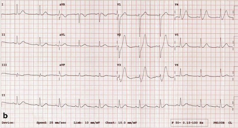
A healthy 58-year-old malepresented to the emergency department (ED) with chest pain, which had started several minutes ago. He was haemodynamically stable with no clinical evidence of heart failure. The first electrocardiogram (ECG) taken 6 min after arrival showed ST-segment elevation in leads I, aVL and V2–6, with ST depression and T wave inversion in lead III, suggestive of an acute anterolateral myocardial infarction (MI). He was treated with aspirin, ticagrelor, nitroglycerin sprays, intravenous morphine and metoclopramide in the ED with relief of chest pain. The initial high-sensitivity cardiac troponin-T level was not elevated. The second ECG taken 28 min after arrival revealed upsloping ST-segment depression > 1 mm at the J point and tall, symmetrical T wave in leads V1–V4. The patient underwent transradial coronary arteriography 55 min after the second ECG, which showed total occlusion of the mid left anterior descending artery (LAD) with thrombolysis in myocardial infarction grade 0 coronary flow and no collateral filling (Rentrop class 0). Thrombectomy, followed by insertion of a drug-eluting stent, was performed uneventfully.
Q. What is the most Probable diagnosis?
Discussion
The de Winter ECG pattern, characterized by 1–3-mm upsloping ST-segment depression at the J point in leads V1 to V6 that continues to tall positive symmetrical T waves, is identified in only 2% of the patients with acute MI. It is regarded as a ‘ST-elevation myocardial infarction (STEMI) equivalent’ because it is highly predictive for acute LAD occlusion. The case is interesting because the ST-segment elevation in the presenting ECG evolved spontaneously to the de Winter pattern before any coronary intervention, indicating that such an evolution does not necessarily coincide with reperfusion. This case demonstrates that the de Winter pattern might be a temporary phenomenon in the trajectory of STEMI. Should there have been any delay in patient presentation or ECG recording, the de Winter pattern could have been the first ECG feature in our case. Given the stable haemodynamic status, absence of heart failure, relief of chest pain after treatment, and a normal initial troponin assay, many clinicians might have treated the patient with ST depression in the presenting ECG as unstable angina conservatively, especially during night time, weekends and holidays when access to cardiac catheterization is less readily available. In that case, the opportunity of treating the total LAD occlusion with primary PCI could have been missed. Therefore, it is important for emergency physicians to recognize the de Winter pattern as a STEMI equivalent and proceed to the cardiac catheterization laboratory without delay.
Intern Emerg Med. 2019. doi: 10.1007/s11739-018-02013-z. [Ahead of print]
Case 4
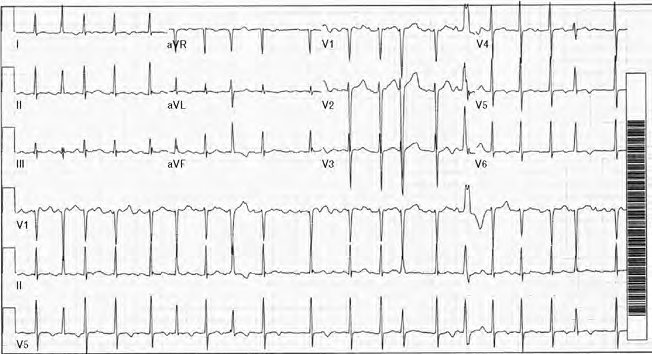
A 45-year-old African–American male came to the emergency room with chief complaints of sudden onset of shortness of breath, chest pain, palpitations, dizziness and sweating that started 1 h prior to presentation. The chest pain was 8/10 in intensity, sudden in onset, stabbing in nature, not related to exertion, with no aggravating or relieving factors. The patient’s past medical history was pertinent for well controlled hypertension and diabetes and several previous ER admissions for recurrent episodes of palpitations, each lasting 20–30 min over the past 2 years. On each of his previous presentations, he was diagnosed with atrial tachycardia of unknown origin and was discharged on atenolol and flecainide. ECG showed atrial fibrillation with rapid ventricular response and aberrantly conducted complexes. Echocardiography done at this presentation revealed a normal left ventricular size with mildly increased left ventricular wall thickness; however, diffuse left ventricular hypokinesis with ejection fraction of 10–20% was noted. He was started on prednisone 1 mg/kg. At the end of 1 month, echocardiography showed improvement in systolic function with ejection fraction improving to 30%.
Q. What is the most Probable diagnosis?
Discussion
Cardiac sarcoidosis is an unusually rare form of sarcoidosis that often remains silent clinically; although if not screened for and diagnosed early can lead to disastrous consequences, which include sudden death. In addition to sudden death, cardiac sarcoidosis can present with myriad manifestations, including congestive heart failure and conduction blocks, as well as atrial or ventricular arrhythmias. Clinical features of cardiac sarcoidosis depend on the location and the extent of myocardium involved. The left ventricular free wall is the most commonly involved, followed by the intraventricular septum. Nearly 70% of patients with cardiac sarcoidosis have ECG abnormalities. Most common findings in ECG include first-degree, second-degree or complete (A–V) block and nonspecific T-wave abnormalities. ECG in conjunction with Holter monitoring is a simple, noninvasive method for screening patients with cardiac sarcoidosis. This is one of the rare instances in which atrial fibrillation was the initial clinical manifestation of sarcoidosis as compared to pulmonary manifestations, which are the presenting features in most cases. Thus, cardiac sarcoidosis should always be kept in the differential diagnosis of atrial fibrillation of unknown origin.
J Cardiovasc Med (Hagerstown). 2015 Jan;16 Suppl 2:S104-12.