Tips and Tricks in ECG Diagnosis
Case 1 - Meningococcal Myocarditis
A 55-year-old woman presented with heavy central chest pain, associated with dyspnea and hypotension, which occurred following 24 hours of self-measured fever up to 39 °C, vomiting, and diarrhea and a 4-day prodrome of sore throat, myalgias, and lethargy. On clinical examination, blood pressure was 72/38mmHg, heart rate was 96 bpm, and temperature was 35.9 °C was recorded. Initial troponin was 72 ng/L, white cell count was 4.34 × 10⁹/ L, and C-reactive protein was 346 mg/L. Screening bloods included Epstein–Barr virus (EBV) serology which demonstrated positive EBV IgM, positive EBV IgG, and positive Epstein-Barr virus nuclear antigen 1 (EBNA) IgG suggestive of recovery or reactivation but not acute primary infection with positive EBNA IgG not likely to appear positive until at least 6weeks after infection. On admission, Electrocardiogram (ECG) showed concave upward diffuse ST-segment elevation (leads II, III, aVF, V2–6) without reciprocal changes and with associated PR-interval depression, not consistent with a single coronary vessel territory.
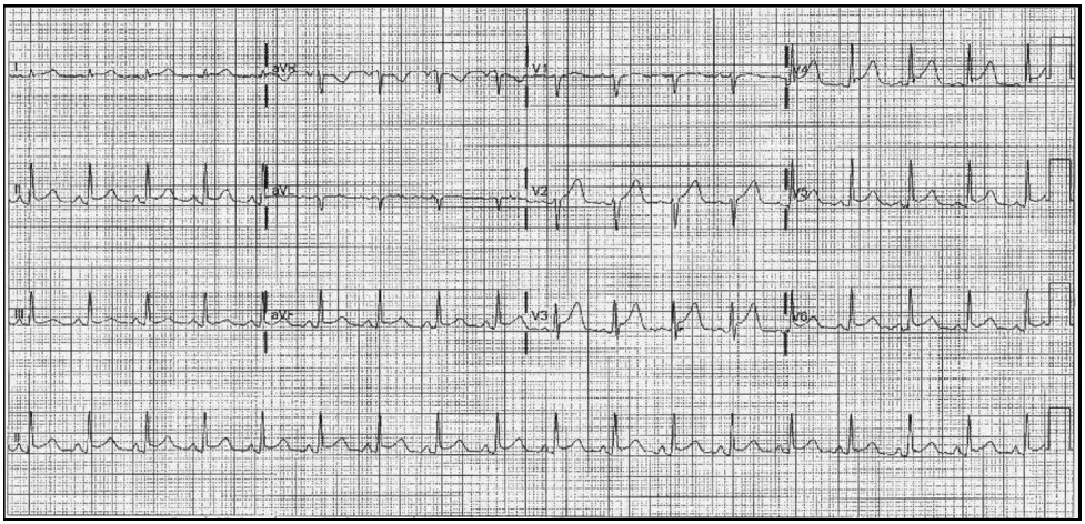
Q. What is the most probable diagnosis?
Discussion
Cardiac involvement can occur following bacteremia secondary to Neisseria meningitides and is likely underrecognized. However, meningococcal disease presenting as primary myopericarditis in immune competent adults is a relatively rare phenomenon. N. meningitidis is a gram-negative diplococcus most commonly responsible for meningococcal meningitis and meningococcemia, both of which may have cardiac manifestations including pericarditis and myocarditis. Primary meningococcal pericarditis is uncommon but well recognized, whereas meningococcal bacteremia presenting with primary myopericarditis is rare. Serotypes causing myopericarditis have included serotype C, serotype Y and serotype W and may occur with or without heart failure. Meningococcal myocarditis is diagnosed through a combination of clinical presentation, diffuse repolarization changes on ECG, and positive blood cultures for N. meningitides. Transthoracic echocardiography has been used in isolated meningococcal pericarditis to help diagnose and guide management. Recently cardiac MRI has emerged as the investigation of choice for nonischemicmyocardial injury to assess heart function and structure. N. meningitidis infection presenting with myopericarditis remains uncommon. Cardiac MRI is the investigation of choice for myocarditis, and patients with myocarditis.
Diagn Microbiol Infect Dis. 2018 Nov;92(3):241-244
Case 2 - Left Ventricular Noncompaction (LVNC)
A 23-year-old female presented for cardiac evaluation because of ECG abnormalities observed during a routine check-up. Physical examination was normal, except for a grade III/VI systolic murmur heard over the apex, radiating to the axilla. Routine blood work was normal, except for an increased NT-proBNP of 3000 pg/mL (normal range 0-125 pg/mL). On the chest x-ray, it showed cardiomegaly. Echocardiography showed left ventricular (LV) enlargement, severe systolic dysfunction with a LV ejection fraction of 25%, restrictive diastolic dysfunction and diffuse LV hypokinesia. LV apical and lateral wall trabeculations were detected. Holter monitoring revealed multiple episodes of unsustained ventricular tachycardia. ECG showed poor R wave progression in leads V1-V3 and negative T waves in leads DI, DII, aVF, V4-V6.
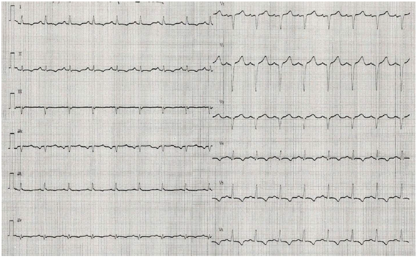
Q. Most probable diagnosis?
Discussion
Left ventricular noncompaction (LVNC) is a rare structural abnormality of the left ventricular myocardium characterized by a spongy endocardial layer and a compacted epicardial layer. Morphologically, the left ventricle (LV) is characterized by the presence of a noncompacted (NC) subendocardial layer, with trabeculae separated by deep recesses, and a thin compacted (C) epicardial layer. The inferolateral and apical regions of the LV are frequently affected. Clinical heterogeneity ranges from asymptomatic to severe or life-threatening manifestations such as heart failure, ventricular arrhythmias or systemic thromboembolism. There is the risk of over diagnosing the condition because several physiological circumstances characterized by volume overload, such as pregnancy, athleticism, African ethnicity, and pathological condition of volume overload, are oftenassociated with a LVNC-like phenotype. LVNC can occur either isolated, or associated with neuromuscular disorders or congenital heart defects. LVNC can frequently be found in primary (dilated, hypertrophic, arrhythmogenic, and restrictive cardiomyopathy) as well as in acquired cardiomyopathies. Even congenital heart diseases are often characterized by ventricular noncompaction.
Rom J Intern Med. 2019 Mar 1;57(1):78-82
Heart Fail Rev. 2019 May;24(3):315-323
Case 3 - Non-ST segment elevation Acute Myocardial Infarction
A 37-year-old man with a history of hypertension and smoking habits was admitted to the emergency room after a self-limited, non-exercise related oppressive chest pain episode. He reported no previous similar episodes and denied accompanying fever or weight loss. He had a history of familial amyloidotic polyneuropathy, being an asymptomatic carrier of an heterozygous mutation in the transthyretin gene on chromosome 18. Blood pressure was 118/81 mm Hg, heart rate was 88 beats per minute, cardiac auscultation revealed normal heart sounds and the absence of heart murmurs. Laboratory findings showed positive high sensitivity cardiac troponin T (hs-cTnT) of 89 ng/L (99th percentile of a reference healthy population upper limit of hs-cTnT 14 ng/L). The 12-lead ECG showed normal sinus rhythm, an early repolarisation pattern but no signs of ischemia.
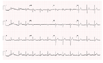
Q. What is the most Probable diagnosis?
Discussion
Non-ST segment elevation acute myocardial infarction (MI) is usually caused by atherosclerotic plaque instability, non-occlusive thrombus formation and distal coronary embolisation. Noncoronary injury to the heart such as cardiac contusion, myocarditis, or presence of cardiotoxic substances can also produce NSTEMI. Finally, conditions relatively unrelated to the coronary arteries or myocardium itself such as hypotension, hypertension, tachycardia, aortic stenosis, and pulmonary embolism lead to NSTEMI because the increased oxygen demand cannot be met. The median age at the time of presentation for ACS in the United States is 68 years. Males outnumber females by 3:2 ratios. The “typical” presentation of NSTEMI is a pressure-like substernal pain, occurring at rest or with minimal exertion. The pain generally lasts more than 10 minutes and may radiate to arm, the neck, or the jaw. The pain may be associated with dyspnea, nausea or vomiting, syncope, fatigue, or diaphoresis. Sudden onset of unexplained dyspnea with or without associated symptoms is also a common presentation. Risk factors for ACS include male sex, older age, family history of coronary artery disease, diabetes, personal history of coronary artery disease, and renal insufficiency. Atypical symptoms may include a stabbing or pleuritic pain, epigastric or abdominal pain, indigestion, and isolated dyspnea. A normal ECG does not exclude ACS and NSTEMI. ST elevation or anterior ST depression should be considered a STEMI until proven otherwise and treated as such. Findings for NSTEMI include transient ST elevation, ST depression, or new T wave inversions. ECG should be repeated at predetermined intervals or if symptoms return. NSTEMI is diagnosed in patients determined to have symptoms consistent with ACS and troponin elevation but without ECG changes consistent with STEMI.
BMJ Case Rep. 2018 Dec 3;11(1).
Non ST Segment Elevation (NSTEMI) Myocardial Infarction. 2019 Feb 28. StatPearls [Internet].
Case 4 - Cardiac Cephalalgia
A 64-year-old man presented with a 6-month history of new headaches. He had a history of migraine and visual aura. His new headaches differed from his typical migraine. Often the headache started with a warm feeling that spread across his chest and up into his neck. This was quickly followed by a generalized headache that reached maximum intensity after a few minutes. The pain was ‘compressing’, and the intensity was severe and debilitating. There were no associated autonomic, visual or neurological symptoms. There were no cardiovascular symptoms during attacks, and in particular no chest pain, dyspnea or diaphoresis. The headaches would last 5–15 min and spontaneously resolve. Over time the attack frequency increased from 2 to 3 times per month to 2–3 times per week. An ECG during one attack showed ST elevation in the inferior leads (II, III and aVF) with reciprocal lateral lead ST depression. A repeat ECG, when headache free, showed spontaneous resolution of these changes.
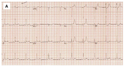
Q. What is the most Probable diagnosis?
Discussion
Cardiac cephalalgia is a migraine-like headache that occurs during episodes of myocardial ischaemia. Clinical characteristics of the headache vary widely but are often severe in intensity, worsen with reduced myocardial perfusion and resolve with reperfusion. It can present along with typical symptoms of angina pectoris, although not always. Cardiac cephalalgia is often aggravated by exercise and improving with rest or nitroglycerine. The diagnosis can be difficult because (as here) the headache onset is not always with exertion, and may occur at rest without chest pain or other cardiovascular symptoms. Headache of cardiac cephalalgia is relatively featureless. Cardiac cephalalgia may be the only symptom of cardiac ischaemia and its complications could be life threatening. Men aged over 60 with risk factors for vascular disease are appearing to be at greatest risk. Cardiac cephalalgia should therefore be considered in patients over 50 years old, with a new (or different), exertional headache, particularly in the presence of risk factors for vascular disease or those experiencing concomitant angina pectoris.
Pract Neurol. 2019 Apr;19(2):173-175.