Tips and Tricks in ECG Diagnosis
Case 1 - Hypertrophic Cardiomyopathy (HCM)
A 37-year-old male patient presented to the hospital with a recent history of decrease in exercise performance since a few months, and also a decrease in exercise heart rate of ~20 beat/ min recorded on a heart rate monitor. Neither personal history of pre syncope and/or syncope nor family history of syncope or sudden death were noted. Physical examination was normal; blood pressure 130/70 mmHg and BMI 23 kg/m2. 12-lead ECG revealed a bi-fascicular block with complete right bundle branch block + left anterior fascicular block (QRS 170 msec), associated to 1st degree AV block with PR interval 250 msec. End-diastolic thickness of septum was 18 mm, posterior and lateral walls 10 mm, and apex 11 mm. There was no systolic anterior motion of the mitral valve and no LV outflow tract gradient was detected either at rest or during exercise
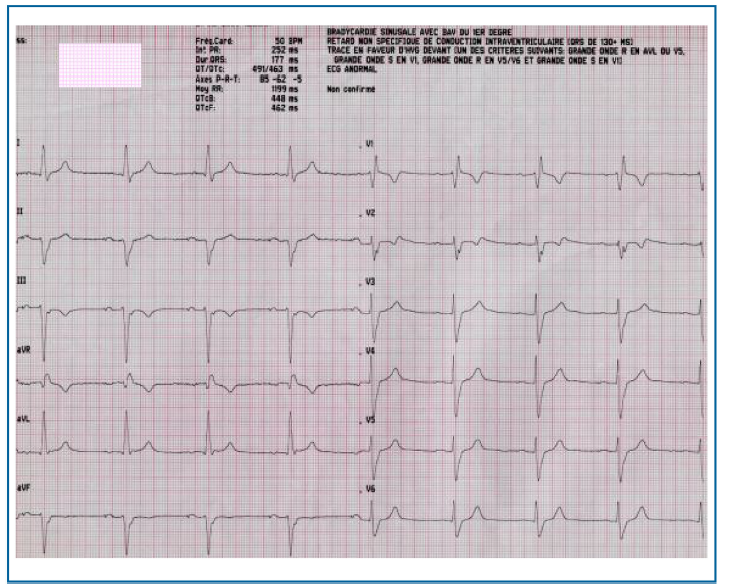
Q. What is the most probable diagnosis?
Discussion
Hypertrophic Cardiomyopathy (HCM) is the most common genetic cardiac disorder. It is characterized by left ventricular (LV) hypertrophy in the absence of another cause for the increased cardiac mass. Prolonged QRS duration and abnormal His-Purkinje system conduction may result in complete AV block. Abnormal His-Purkinje conduction in 23% of HCM patients who survived sudden cardiac death. The identification of patients who are at risk for sudden death remains an important challenge and emerging role of cardiovascular magnetic resonance imaging (MRI) for risk stratification is increasingly highlighted. The Pathophysiology of AV block in human HCM is unknown. In human's with complete AV block, it has been generally assumed that the wide escape-beat QRS complexes may be attributed to lesions in the distal portion of the AV bundle or bilateral lesions of the bundle branches, and that the narrow QRS complexes may be associated with lesions in the approaches to the AV node, in the AV node itself, or in the proximal portion of the AV bundle.
Presse Med. 2019 Jan;48(1 Pt 1):68-71
Case 2 - Iatrogenic Takotsubo Cardiomyopathy
A 50-year-old woman presented to the emergency department with complaints of lip and tongue swelling and shortness of breath after ingestion of fast food, which she has consumed without issue in the past. Pulse oximetry was noted to be 100% on room air. She was found to be oxygenating well, speaking full sentences. She was noted to have pallor, diaphoresis, and central cyanosis. Her blood pressure rate was 208/136. The patient complained of nausea and crushing substernal chest pain. Electrocardiogram was initially deemed to be an ST-segment elevation myocardial infarction but officially read as a sinus rhythm with first- and second-degree atrioventricular block, competing accelerated idioventricular rhythms and a fusion beat. Serum troponin I levels were elevated at 2.54 ng/mL, with repeat at 3.77 ng/mL.
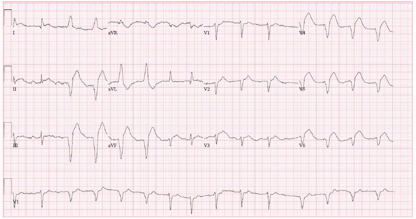
Q. Most probable diagnosis?
Discussion
Takotsubo Cardiomyopathy (also known as stress Cardiomyopathy) is a reversible systolic dysfunction because of myocyte injury precipitated by significantly elevated serum catecholamine levels, typically from endogenous sources released during acute emotional or physical stress. The apical ballooning seen on echocardiography and cardiac catheterization resembles a takotsubo, or Japanese octopus pot, from which the syndrome receives its name. Stress cardiomyopathy results from catecholamine excess. Several lines of evidence support this mechanism. Plasma catecholamine levels are elevated in patients with stress Cardiomyopathy. Stress cardiomyopathy is a neurocardiogenic phenomenon—it can be precipitated by emotional stress, other stress states, drugs, particularly catecholamines, or neurologic injury, although lack of an apparent external stressor does not rule out the diagnosis. Most cases present with chest pain and/or dyspnea, and occur in post-menopausal women, for reasons that remain uncertain. The ECG is abnormal, with ST elevation in 30–50 % of patients, and precordial T wave inversions, often dramatic, in most of the rest.
Am J Ther. 2018 Nov/Dec;25(6):e695-e698.
Intensive Care Med.2016 Mar;42(3):432-435
Case 3 - ST segment elevation myocardial infarction
A 50-year-old man collapsed at the roadside with retrosternal pain, shortness of breath and generalized weakness. He appeared pale and clammy and was confused. Systolic blood pressure was 90 mm Hg in the right arm. He was a smoker and had a history of hypertension. The ECG on arrival to the emergency department was reported as demonstrating ST segment elevation of up to 1.5 mm in leads V1 to V3, leading to a diagnosis of an acute ST-elevation myocardial infarction. The patient was loaded with oral antiplatelet therapy and immediately transferred to the cardiac catheterization laboratory for further investigation. Following a 20-day hospital admission, and postoperative atrial fibrillation, the patient made a steady and full recovery. He was anticoagulated, with a recommendation to titrate antihypertensive medication to maintain blood pressure at 120/70.
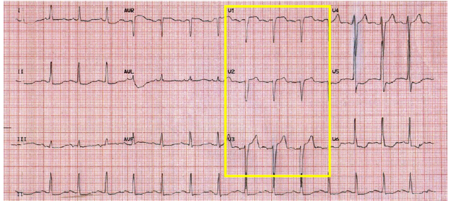
Q. What is the most Probable diagnosis?
Discussion
ST-segment elevation myocardial infarction or STEMI continues to be one of the most dangerous acute complications of coronary artery disease. Anterior ST-elevation myocardial infarction is rare but can occur due to compression of coronary Ostia by hematoma, or occlusion by extension of the dissection flap. patients with STEMI undergo rapid revascularization, most commonly with primary Percutaneous coronary intervention. The causes of death were cardiovascular in 70% of patients with obstructive coronary artery disease, 38% in those with non obstructive disease, and 32% in those with normal coronary arteries. ST-segment elevation acute coronary syndrome patients with non-obstructive coronary artery disease had a long term risk of death similar to or higher than that of patients with obstructive disease. A 12-lead ECG is important in differentiating different heart condition, but clinicians should be mindful that presence of left ventricular hypertrophy can be misleading. ST-elevation acute coronary syndrome patients without obstructive coronary artery disease warrant medical attention and close follow-up.
BMJ Case Rep.2019 Jan 14;12(1).
Eur Heart j. 2018 Jan 7;39(2):75-78.
Case 4 - Wellens’ Syndrome in pulmonary Embolism
A 22 year old man presented to the emergency room with the sudden onset of intermittent left-sided chest pain and dyspnoea on exertion for 1 day. The patient was in a usual state of health until 1 day before presentation. Pain was described as cramping in type with no radiation and referral. He denied history of fever, sick contacts, nausea, vomiting, palpitation and light-headedness. He denied history of alcohol, tobacco, illicit drug and cocaine use. Family history of heart disease and clotting disorder was negative. His blood pressure was 118/70 mm Hg, heart rate was 78 beats/min in sinus rhythm, respiratory rate was 14 breaths/min and saturated 100% on room air. Twelve lead ECG during pain-free interval revealed biphasic T waves in leads V2 and V3. ECG revealed sinus rhythm, normal progression or R waves in precordial leads, isoelectric ST segment in the absence of pathological Q waves.
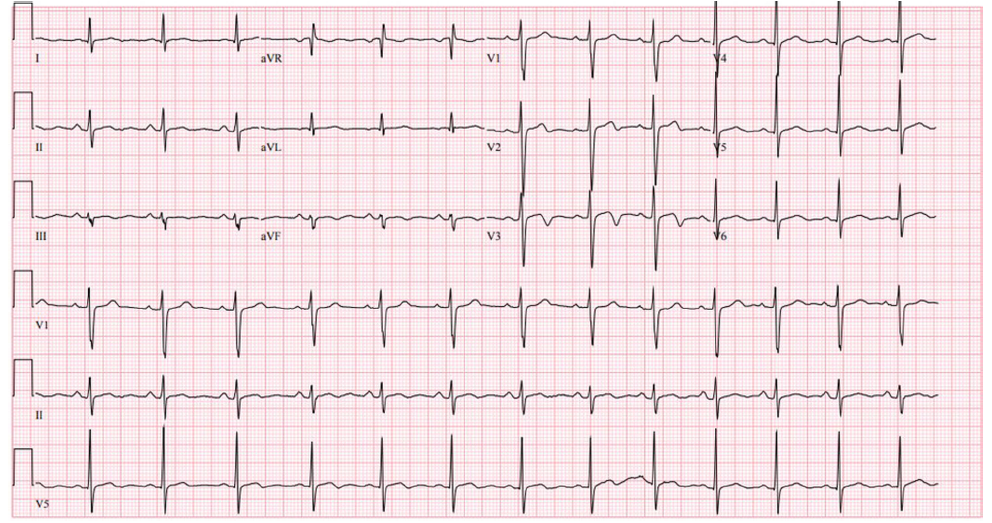
Q. What is the most Probable diagnosis?
Discussion
Wellens’ syndrome is described as characteristic biphasic or symmetrical T-wave inversion with normal precordial R-wave progression and the absence of Q waves in the right precordial leads. Wellens’ syndrome was first described as a characteristic T-wave changes in precordial leads in a subset of patients with unstable angina. Wellens’ syndrome has two distinct morphological subtypes. The more common type A is described as symmetrical T-wave inversion in V2 and V3, often in V4 and V5 and sometimes in V6.Type B is less common and presents with biphasic T waves in leads V2 and V3 as noted in our patient. Type B Wellens’ syndrome is associated with a relatively higher mortality. Wellens’ syndrome is well known in cardiology as a subtle yet ominous manifestation of critical proximal LAD occlusion. Biphasic T waves have also been reported in association with coronary vasospasm,4 and infusion-related hypersensitivity to daratumumab. This Case further highlights the importance of recognizing this devious sign as it may be a potential manifestation of other alternative life-threatening condition like PE with right ventricular strain. Wellens’ ECG change was seen in our patient in the setting of pulmonary embolism (PE) with right ventricular strain. T-wave inversion in right precordial leads V1-V3 is a well-recognized subtle ECG change in right ventricular strain; however, biphasic T waves in the setting of PE.
BMJ Case Rep. 2018 Dec 19;11(1)