A 58-year-old man was admitted to the hospital with complaints of acute onset of confusion, agitation,
and sweating.
The patient had medical history of severe major depressive disorder and attention deficit hyperactivity
disorder (ADHD).
Investigations
Initial examination was remarkable for fever of 38.1°C, sinus tachycardia, tachypnea, upper chest and
face flushing, altered mental status, hyperreflexia, rigidity, and resting fine tremors.
Then, the patient had an episode of behavioral arrest followed by rhythmic clonic movements of the left
upper extremity for 3–4 min. The patient was hard to arouse for a few hours afterward.
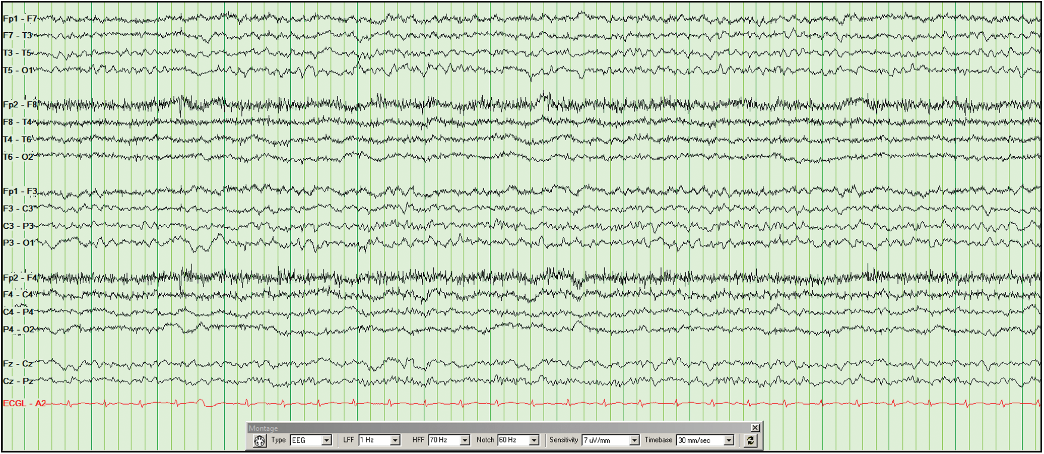
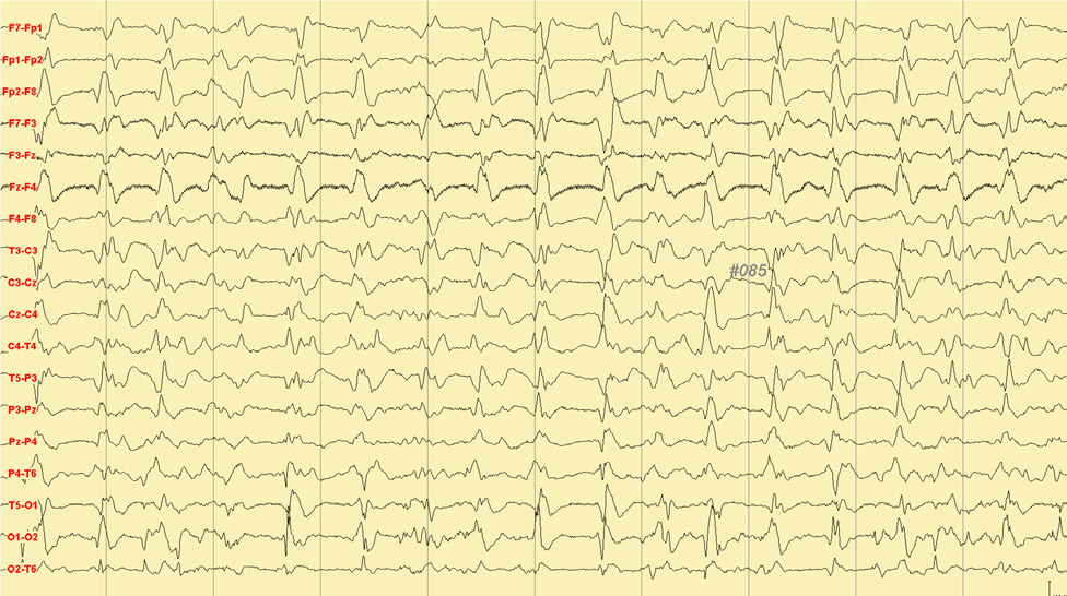
The Electroencephalography (EEG) was performed and was remarkable for continuous focal slowing over the
right temporal region.
The patient was started on anti-seizure medication (levetiracetam).
The patient was placed on continuous EEG monitoring, which initially showed right side temporal mixed
delta and theta focal slowing in addition to asymmetric decreased amplitude over the right side (Figure
1).
The patient was started on standing benzodiazepine doses and cyproheptadine.
His symptoms gradually improved and a routine EEG study was carried out and was only significant for
mild generalized slowing, without any focal findings.
Figure 1: Electroencephalogram shows right side temporal mixed delta and theta focal slowing in
addition to asymmetric decreased amplitude over the right side on a longitudinal bipolar montage
Diagnosis
The patient was diagnosed with serotonin syndrome.
Discussion
Serotonin syndrome is a life-threatening condition. Neuromuscular excitation, autonomic nervous system
hyperactivity, and change in mental state are the core manifestations of this syndrome. Seizures related to
serotonin syndrome can be secondary to hyperthermia that exceeds 40°C, electrolyte abnormalities, specific
medication combinations and specific medication overdosing. It is most commonly caused by combining two or
more serotonergic agents, which can include drugs for many different purposes (antibiotics, antiemetics,
supplements, etc.), not just psychiatric agents. Recognition of seizure as a symptom of serotonin syndrome
is very important for early treatment and avoidance of long-term consequences. The suggested pathophysiology
for seizures in the setting of serotonin syndrome is likely related to cortical excitement in the prefrontal
region given that it is rich with 5HT receptors in animal modules. Treatment of serotonin syndrome is mostly
supportive.
EEG Tips:
EEG findings help to differentiate serotonin syndrome from other acute causes of seizures, especially
neuroleptic malignant syndrome.
2. Acute visual impairment as a symptom of non-convulsive status epilepticus
Case presentation
A 65-year-old man was admitted to the neurology department complaining of an inability to see in the
right eye for 2 days along with acute headache.
He had a history of atrial fibrillation in rheumatic heart disease and suffered an ischaemic stroke in
the left frontotemporal parietal lobe 4 years ago.
Two years ago, he suffered symptomatic epilepsy due to ischaemic stroke and effective antiepileptic
drugs were prescribed.
Investigations:
At the first medical evaluation, the arterial pressure was 170/100 mmHg, and arterial blood lactic acid
was 3.3 mmol/L.
Liver function, renal function, electrolytes, erythrocyte sedimentation rate, procalcitonin,
cerebrospinal fluid examination, syphilis antibody and HIV testing were normal.
Neurological examination revealed a cognitive decline wherein the patient could not accurately answer
questions or simply reply with short sentences or words.
On Ophthalmological examination it was found that neither eye of the patient could read a visual chart
clearly, but he could grasp a pen when shaken in front of eyes, suggesting macular avoidance or partial
visual field avoidance.
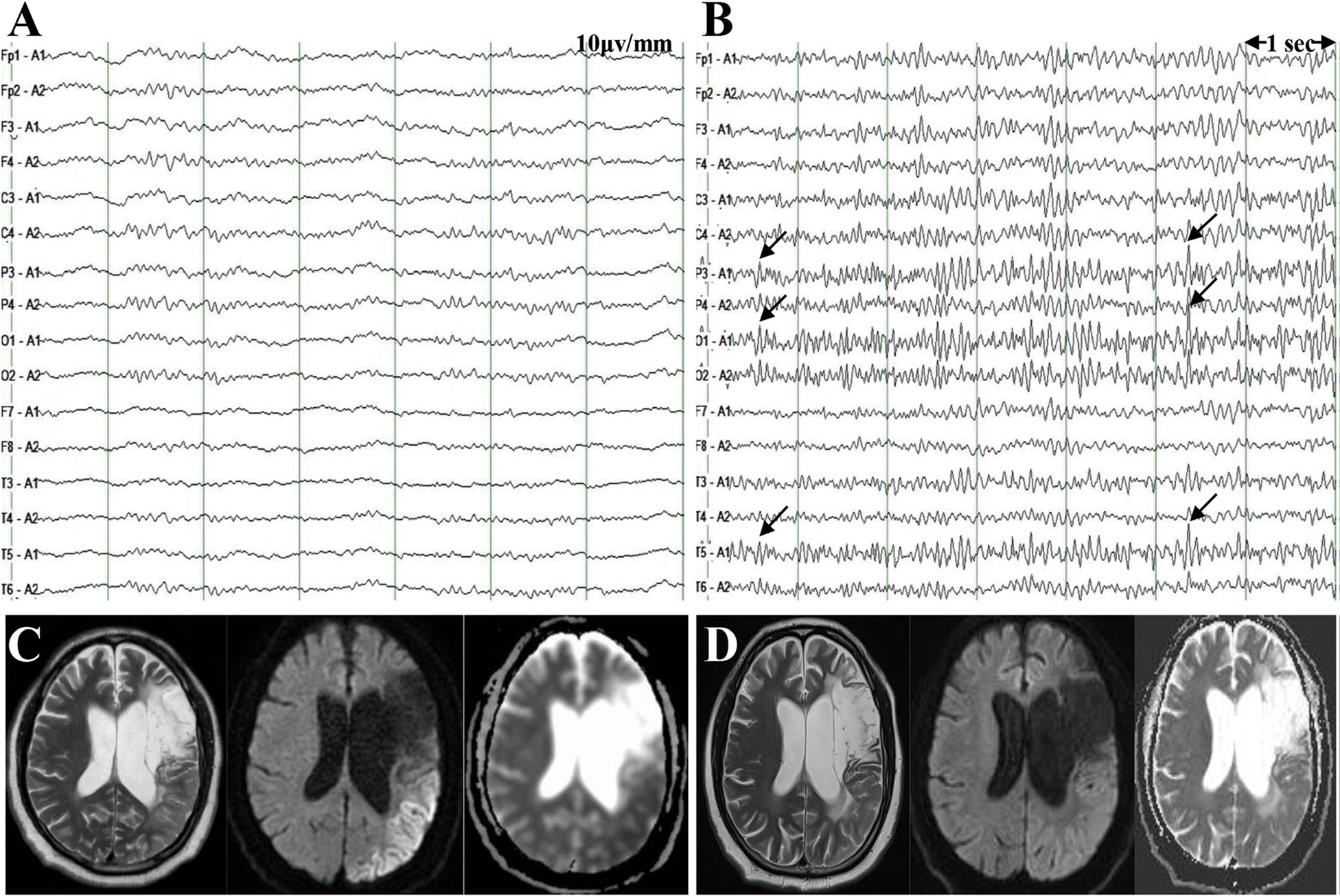
An MRI was then performed, which showed marked diffuse cortical hyperintensities in the left
temporoparietal and occipital lobes on T2 and diffusion-weighted images.
EEG monitoring was performed, which found continuous sharp wave discharges, especially in the regions of
the left temporal, parietal and occipital lobes (Figure 1).
Figure 1: Ictal EEG showed continuous sharp wave discharges mainly in the regions of the left
temporal, parietal and occipital lobe
Diagnosis
The patient was diagnosed with Nonconvulsive status epilepticus (NCSE)
Discussion
Nonconvulsive status epilepticus (NCSE) is a state of ongoing seizure activity without convulsions. NCSE
represents a major problem in neurological critical care patients. The variable and subtle clinical symptoms
of NCSE, such as staring, repetitive blinking, chewing, swallowing, automatism, altered mental status,
subtle facial or limb twitches, speech disturbance and extrapyramidal signs, make diagnosis and treatment
challenging. For patients with atypical symptoms of NCSE, it is very important that the diagnosis of NCSE
meet the EEG criteria. In NCSE, the activated cortex is in an extreme electrophysiological state that
exhibits increased glucose and oxygen usage, thereby causing compensatory regional hyperperfusion.
Progressive dementia, acute visual impairment could be another clinical symptom of NCSE if the occipital
cortex is involved. Acute visual impairment can present as a main symptom of NCSE when the temporoparietal
occipital lobes are involved, and its possible mechanism may be hemianopsia and visual neglect.
EEG Tips:
Electroencephalography (EEG) monitoring has been associated with an early diagnosis of Nonconvulsive
status epilepticus (NCSE).
3. Epileptic encephalopathy with continuous spike-and-wave during sleep
Case presentation:
A 10-year old girl was admitted with complaints of frequently recurring generalized tonic and myoclonic
seizures.
She was the second child of consanguineous healthy parents, without a familial history of epilepsy.
She was born full term after a normal pregnancy.
Investigations
On physical examination, the patient has the following growth parameters; 16 kg, height 115 cm and head
circumference 43 cm.
She has generalized muscular hypotonia.
She has no head control, and she is not capable of dependent sitting.
She was not able to neither follow instructions nor speak single words.
On the 20th day, she had recurrent, afebrile focal myoclonic and clonic seizures.
Cranial magnetic resonance imaging (MRI) at the age of 2 months and 3 years were normal.
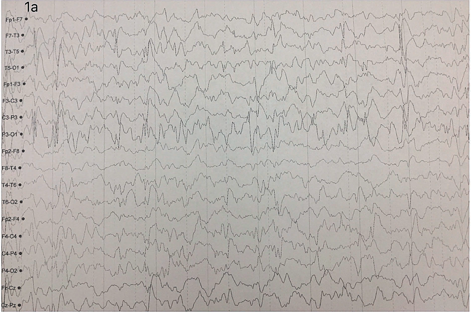
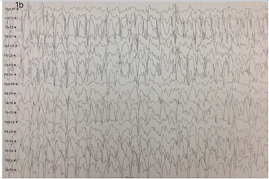
Interictal EEG revealed generalized spike and slow-wave activity, occurring intermittently in
wakefulness but present for at least 85% of non-REM sleeps [Figure 1 (a) and (b)].
• Interictal EEG revealed generalized spike and slow-wave activity, occurring intermittently in
wakefulness but present for at least 85% of non-REM sleeps [Figure 1 (a) and (b)].
Diagnosis
The patient was diagnosed with cyclin-dependent kinase-like 5 (CDKL5) disorder.
Discussion
Mutations of the X-linked gene cyclin-dependent kinase-like 5 (CDKL5) have been associated with early
infantile epileptic encephalopathy 2 and early-onset seizures and symptoms sharing features of atypical Rett
syndrome. CDKL5 mutations reported predominantly in females. The patients with the CDKL5 disorder have
multiple types of seizures: epileptic spasms, tonic seizures, absences, and myoclonic seizures. Interictal
electroencephalography (EEG) is often normal at early stages however it shows the progressive deterioration
of the background rhythm during the disease course. CDKL5 mutated patients tend to present early seizures
with normal interictal EEG then infantile spasms with hypsarrhythmia, and finally multifocal and refractory
epilepsy.
EEG Tips:
Epileptic encephalopathy with continuous spike-and-wave during sleep is an EEG pattern usually
age-related and occurs commonly during the childhood.
4. New-onset refractory status epilepticus secondary to antiphospholipid syndrome
Case presentation:
A 43-year-old man was admitted with complaints of blurry vision and headache.
He was a non-smoker and had no relevant family or medical history.
He developed generalized tonic-clonic seizure after 2 hours of admission.
Investigations:
The patient’s workup findings of CT scan and brain MRI were normal.
The lumbar puncture which only showed slightly elevated white blood cells with normal protein and
glucose levels.
After 2 weeks, he was comatose and was having intermittent twitching of the face and upper extremity.
Head CT, MRI and neck magnetic resonance angiography and venography (MRA/MRV), in addition to cerebral
angiography showed no abnormality detected except for a parasagittal hematoma.
He had a prolonged seizure that lasted more than 10 minutes.
He continued having in between seizures activities while he was being kept intubated and ventilated on
tracheostomy with ketamine and thiopental.
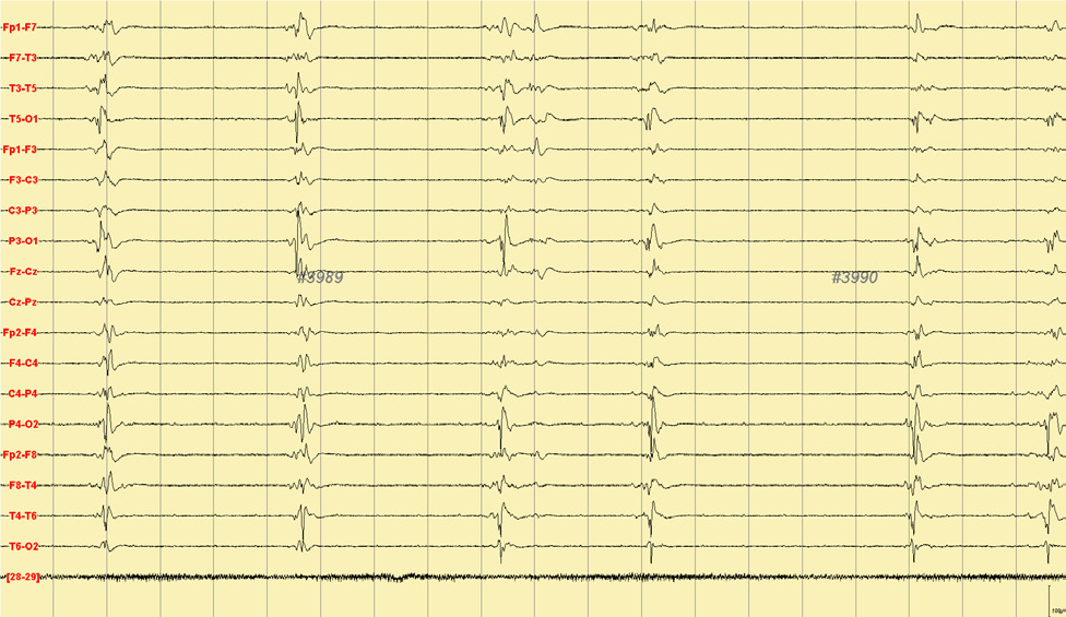
Continuous EEG showed initially generalized spikes and waves every 1-2 seconds for >48 hours (Figure 1).
Then, anesthesia induced burst suppression pattern was observed (Figure 2).
Work-up revealed elevated Antiphospholipid antibody (APA) including antiB2 Glycoprotein I.
Multiple intravenous doses of benzodiazepine and a loading dose of phenytoin, valproic acid,
levetiracetam, midazolam, thiopental, phenobarbitone, fentanyl, and ketamine infusion were administered.
A trial of 5 days induced hypothermia ended up with insufficient seizure suppression.
The patient was diagnosed with New-onset refractory status epilepticus (NORSE).
Discussion
New-onset refractory status epilepticus (NORSE) is a drug-resistant status epilepticus that often has a
catastrophic outcome. A recent study concluded 32% of NORSE patients ended up with altered behavioral states
like aggression. Autoimmune disorders result in various neurological and psychiatric presentations.
Antiphospholipid syndrome (APS), an acquired autoimmune disorder, can manifest a spectrum of clinical
manifestations, including pregnancy morbidities; vasculopathies; and neuropsychiatric disorders such as
stroke, seizures, and psychosis. Mechanism of the pathogenesis of epilepsy secondary to APS could be linked
to the immune-mediated response and vasculopathy inherent to APS. Autoimmune epilepsies are also known to be
linked with refractory status epilepticus, the outcome of which is primarily determined by the underlying
etiology of the seizures, age of the patient and duration of status epilepticus
EEG Tips:
Early psychiatric referral and EEG is recommended and helpful for vigilance of psychological
complications of refractory status epilepticus’ patients.