1. Sudden Death After Febrile Seizure Case Report: Cerebral Suppression Precedes Severe Bradycardia
Case presentation
A 20-month-old girl presented with a complicated medical history underwent prolonged VEM after first presentation of convulsive febrile status epilepticus.
The child’s medical history showed a chromosomal disorder, with bilateral cleft palate, short webbed neck, congenital heart disease, and severe bilateral bronchomalacia.
The girl had a respiratory insufficiency, chronic hypoglycemia, and frequent infections leading to the prolonged hospitalization.
Neurologic disturbances which included hypotonia from birth and over all developmental delay.
MRI at the young stage showed complified gyral pattern, hypo-opercularization of the temporal lobes, and hypoplastic olfactory bulbs and tracts and in discussion with the family intubation or chest compressions were not permitted even in acute deterioration.
At the same age, the child had her first seizure with convulsive febrile status epilepticus which lasted for 45 minutes and was admitted to the hospital.
On day 2, another seizure occurred with hypoglycemia, and the treatment was started with levetiracetam, and on day 3, VEM was started to assess subclinical seizures at 6.5 hours of recording code blue was called for cessation of breathing as the girl died.
Case Investigations
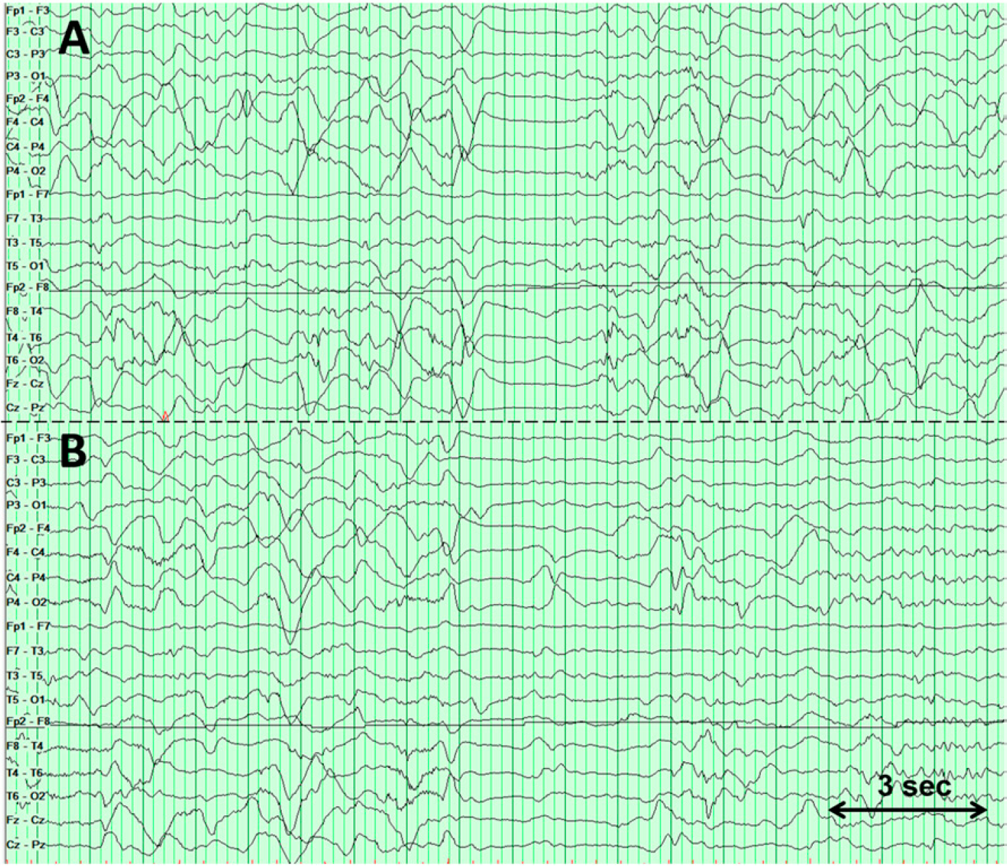
EEG showed diffuse, high-amplitude delta activity, with relative attenuation over the left hemisphere, consistent with a diffuse encephalopathy. Frequent multifocal spikes were observed from the left frontal, right posterior temporal, and left anterior temporal regions. Clinical or electrographic seizures were not seen around 6.5 hours of VEM before death.
ECG showed sinus rhythm for the first 6 hours of recording at T-10 minutes. At T-8 minutes, profound sinus bradycardia was seen with ventricular escape beats.
Heart rate was steady at 120-135 beats/min until T-8 minutes, after that the heart rate decreased and became stable around 70 beats/min.
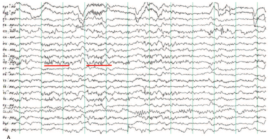
Diffuse cerebral suppression on EEG showed A) During the 6 hours leading up to death; diffuse high-amplitude slow waves are seen, a brief period of generalized attenuation was also observed. B) Ten minutes before terminal asystole, there is an abrupt onset of diffuse cerebral suppression, without any recovery.
Diagnosis
On EEG Diffuse cerebral suppression was observed 10 minutes before death, followed by severe bradycardia and increased heart rate variability.
Discussion
The present study describes the sequence of events leading up to sudden unexpected death in childhood (SUDC), days after initial presentation with febrile status epilepticus. The girl did not meet the criteria for a diagnosis of epilepsy although the EEG suggested a high risk of further seizures. Physiologic monitoring data suggested the cause of death was rooted in the central nervous system, following a pattern reminiscent of recently published adult SUDEP cases. The first observed physiologic change was diffuse cerebral suppression on the EEG, a feature universally reported in adult SUDEP during VEM. In retrospect, the main warning sign for the girl patient was brief periods of diffuse cerebral suppression suggesting cerebral dysfunction was the initial trigger for the cascade of events leading to death.
The diffuse cerebral suppression was followed minutes later by increased parasympathetic activity and this phenomenon is supported by the recent series of Lhatoo et al. Risk of sudden death in children with seizures may be increased if brief periods of generalized EEG suppression are observed. This case shows that children with febrile seizures can have sudden death in a manner reminiscent of adult SUDEP. This case may be representative of the physiologic changes preceding death in pediatric SUDEP; however, this theory cannot be confirmed without identification and investigation of definite SUDEP cases involving children during VEM.
EEG Tips:
EEG showed diffuse, high-amplitude delta activity, with relative attenuation over the left hemisphere, consistent with a diffuse encephalopathy.
2. Intractable ictal vomiting: a new form of reflex epilepsy?
Case presentation
A 21-year-old female college student started experiencing distressing episodes of prolonged nausea, vomiting, and retching five months ago.
An individual bout lasting an hour with severe retching and a constant urge to empty the contents of the stomach.
All of the episodes occurring 1-3 times a day were temporally related to food intake, starting within a few to a maximum of 10 minutes after a meal. There were no other clinical accompaniments, antecedent events, or possible precipitating causes.
The patient studies and social and family life were severely hampered. After 3 months of illness, she started losing consciousness in 1-2 times a month.
Systemic and neurological examination was unremarkable. The patient soon underwent a 32-channel awake and sleep EEG recording of 45 minutes to explore an ictal origin. She was reinitiated on valproate with no more episodes reported ever since.
Clinical investigations:
Video-EEG revealed intermittent frontocentral (F3/C3) discharges and were convincing enough to conclude that the patient was having a form of epilepsy dominated by ictal vomiting. episodes of vomiting and retching.
A brain MRI epilepsy protocol did not reveal any focal pathologies of the brainstem or posterior fossa or any anatomical substrates responsible for epileptogenesis.
Figure1. EEG recording of patient during an episode of vomiting and retching
Diagnosis
Intermittent frontocentral (F3/C3) discharges seen were convincing enough to conclude that the patient was having a form of epilepsy dominated by ictal vomiting.
Discussion
This case report describes a young girl patient with autonomic seizures of ictal vomiting and occasional focal seizures. The report signifies the importance to discuss on the rare and often misdiagnosed ictal expression and its roots. In an analysis of 24 patients with ictal vomiting by Panayiotopoulos, benign childhood epilepsies emerged as the leading cause, especially those of occipital origin. PS is typically seen in the 4-6-year age group. In adults it is originated primarily from the temporal lobe and is usually accompanied by other ictal phenomena. Among the benign occipital lobe epilepsies, Panayiotopolous syndrome and idiopathic photosensitive occipital lobe epilepsy can exhibit ictal vomiting.
This case therefore bears resemblance to the various epilepsies discussed but does not confirm entirely. The responsiveness to valproate points towards a diagnosis of generalized epilepsy but the consistent focality on interictal EEG cannot be overlooked. Finally, it is proposed that this is a unique form of partial epilepsy recruiting a wider network responsive to valproate, resulting in ictal vomiting with a clearly evident reflex component.
EEG Tips:
EEG recording helped confirm epilepsy dominated by ictal vomiting as evidenced by intermittent frontocentral (F3/C3) discharges.
3. De novo 12q22.q23.3 duplication associated with temporal lobe epilepsy
Case presentation:
This girl was the second child of Caucasian healthy, non-consanguineous parents with unremarkable family history. The girl was born at 37 weeks + 5 days gestation by a cesarean section.
At birth, she weighted 3460g (75-90th centile), length was 51 cm and OFC circumference was 34.5 cm.
The girl’s development milestones were slightly delayed with head control at 4 months, sitting position at 10 months, and she started walking at 17 months. She pronounced her first words at the age of 2 years and 5 months.
At 30 months, the patient experienced a febrile tonic-clonic seizure. At age 3 years, she was admitted due to a febrile seizure featuring loss of contact, oromasticatory automatisms, and left head deviation, lasting less than 5 minutes. From the following months, the girl suffered from afebrile weekly dyscognitive seizures with impaired contact and oromasticatory or gestural automatisms, lasting up to a few minutes.
After examination, the patient presented with macrosomy [weight 22,7 kg (>97th centile), height 104 cm (>97th centile), OFC 51 cm (50th centile)].
Also revealed slight dysmorphic features of straight eyebrows, sinophris, abnormal internal canthal folds, large nasal philtrum, small mouth, bilateral shortness of the fifth finger, bilateral syndactyly of the second and third toes and revolved fifth toenails.
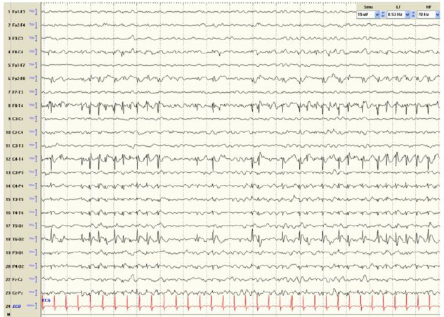
Interictal EEG revealed high-voltage spike and waves complexes over the posterior right temporal lobe areas. Treatment with carbamazepine (30 mg/Kg/day) resulted in complete seizure control and disappearance of EEG abnormalities
1.5 Tesla brain MRI was unremarkable.
Genetic analysis by 44K kit revealed 12 Mb duplication at e12q22-q23.3 which was absent in the girl’s parents.
Figure 1. Interictal EEG recording showing high-voltage spike and waves complexes over the posterior right temporal lobe areas.
Diagnosis
Genetic analysis by 44K kit array-CGH by Agilent Technologies, revealed a 12 Mb duplication at e 12q22-q23.3.
Interictal EEG showed high-voltage spike and wave complexes over the posterior right temporal lobe areas.
Discussion
Temporal lobe epilepsy (TLE) is the most common form of focal epilepsy with acquired central nervous system lesions or genetic. Various susceptibility genes and environmental factors are believed to be involved in the aetiology of TLE, which is considered to be a heterogeneous, polygenic, and complex disorder. Rare point mutations in LGI1, DEPDC5, and RELN as well as some copy number variations (CNVs) have been reported in families with TLE patients.
This is the first report of de novo 12q22-q23.3 duplication associated with TLE. An overlapping de novo duplication (ID 258582) has been reported in the DECIPHER v8.9 database. In this case, the 12Mb duplication at 12q22-q23.3 encompasses genes. Finally, this report confirms that 12q22-q23.3 is a candidate locus for familial TLE with febrile seizures and highlights the role of chromosomal rearrangements in patients with epilepsy and intellectual disability.
EEG Tips:
Interictal EEG helped in displaying high-voltage spike and wave complexes over the posterior right temporal lobe areas.
4. Burst Suppression Pattern on EEG Secondary to Valproic Acid-Induced Hyperammonemic Encephalopathy
Case presentation:
Presented here a 13-year-old girl with intractable epilepsy.
Her medical history was significant for autism and behavioral problems for which she was on risperidone.
She was not on any anti-epileptic medication. At her baseline she was ambulatory and had a few words.
Poor response was noticed after treatment with Valproic acid with dose of 1,575 mg twice/day for 2 weeks.
She was admitted to another hospital and started getting treatment with 10 mg of diazepam rectally followed by 4 mg of lorazepam, 20 mg/kg of fosphenytoin anf finally with 6 mg/kg of fosphenytoin. However, to avoid persistent seizures she was given 10mg/kg of Phenobarbital.
On further examination, she was found to be afebrile and lethargic and was not responsive to verbal stimuli. She moved her head and grimaced to chest rub with her pupils more reactive and had an intact gag reflex.
Case investigations:
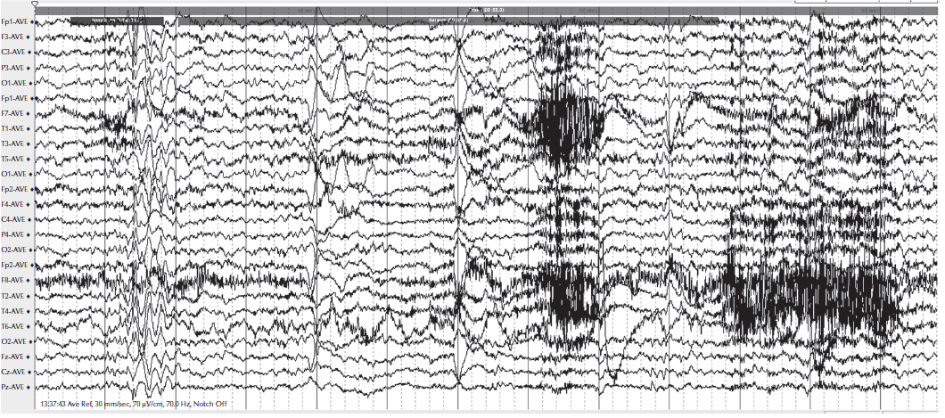
EEG showed a true burst-suppression pattern as shown in Figure. 1, rather than prolonged status epilecticus.
Complete blood count, serum electrolytes, glucose, calcium, and liver enzymes were within normal limits. The ammonia concentration was found to be increased (upto 101 μmol/L). The serum valproic acid level was elevated, at 269.9 mg/L.
Cerebrospinal fluid (CSF) and MRI results were reported to be normal.
Figure 1. EEG recorded on the day of admission, showing a burst-suppression pattern
Diagnosis
EEG showed a burst-suppression pattern confirmed hyperammonemic encephalopathy induced by valproic acid.
Discussion
This is the first report of a burst suppression EEG occurring as a consequence of valproic acid-induced hyperammonemic encephalopathy. Hyperammonemic encephalopathy seems to be more frequent in patients treated with valproate who have carnitine deficiency or congenital enzymatic disturbances like urea cycle defects. In this patient, the symptoms emerged about two weeks after the last increase in the valproic acid dose.
An EEG burst-suppression pattern consisting of an unaltered pattern of periods of low voltage (less than 10 μV) with bursts of higher amplitude activity with frequencies combined with spikes and sharps waves. This EEG pattern was noticed in a range of clinical conditions like in head trauma, stroke, coma, anoxia, anesthesia, hypothermia, and medication overdose as well as in premature babies. The bursts of this patient initially were pleomorphic and non-stereotyped with occasional sharply-contoured waveforms without resembling ictal patterns.
Finally, though there is no relationship between daily dosages and serum concentration of valproic acid, the degree of hyperammonemia and the severity of the encephalopathy. Simultaneous administered anti-seizure medications like phenobarbital, topiramate and rarely with phenytoin and carbamazepine exacerbated valproic acid-induced hyperammonemic encephalopathy. Therefore, valproic acid treatment may cause both the hyperammonemic encephalopathy and the burst-suppression pattern in the EEG.
EEG Tips:
EEG revealed a burst-suppression pattern in a patient of valproic acid-induced hyperammonemic encephalopathy