1. Intractable Epilepsy Resultant of Cortical Development Malformations
Case presentation
A 10 year old right hand dominant boy with medically intractable epilepsy, global developmental delay
and left hemiparesis presented to the clinic for possible surgical evaluation for his seizure disorder.
He had no history of febrile seizures, significant head injury or brain infection.
He delivered by repeat cesarean section at term and received some bag‐mask oxygen at birth.
Investigations
The first seizure onset was at age 3 when he presented with a seizure characterized by jaw clenching,
repeated emesis with eye deviation to the left, which lasted for 2‐3 minutes and was followed by
postictal limpness. He was commenced on valproate.
At eight years of age, seizures again recurred but were unresponsive to higher doses of valproate.
On arrival to the center, he had global developmental delay and had plateaued, although not regressed in
development, since recurrence of his seizures.
He could recognize approximately 14 letters, knew some letter sounds and could rote count.
His examination was significant for a left hemiparesis which was longstanding as suggested by a smaller
left hand and foot and shorter left leg.
He was able to ambulate independently and while his left hand was weaker, he did have some independent
finger movement.
No neurocutaneous lesions were noted on Woods lamp exam.
The remainder of his exam was unremarkable.
His MRI demonstrated an extensive right hemispheric malformation of cortical development most prominent
within the right frontal and occipital lobes.
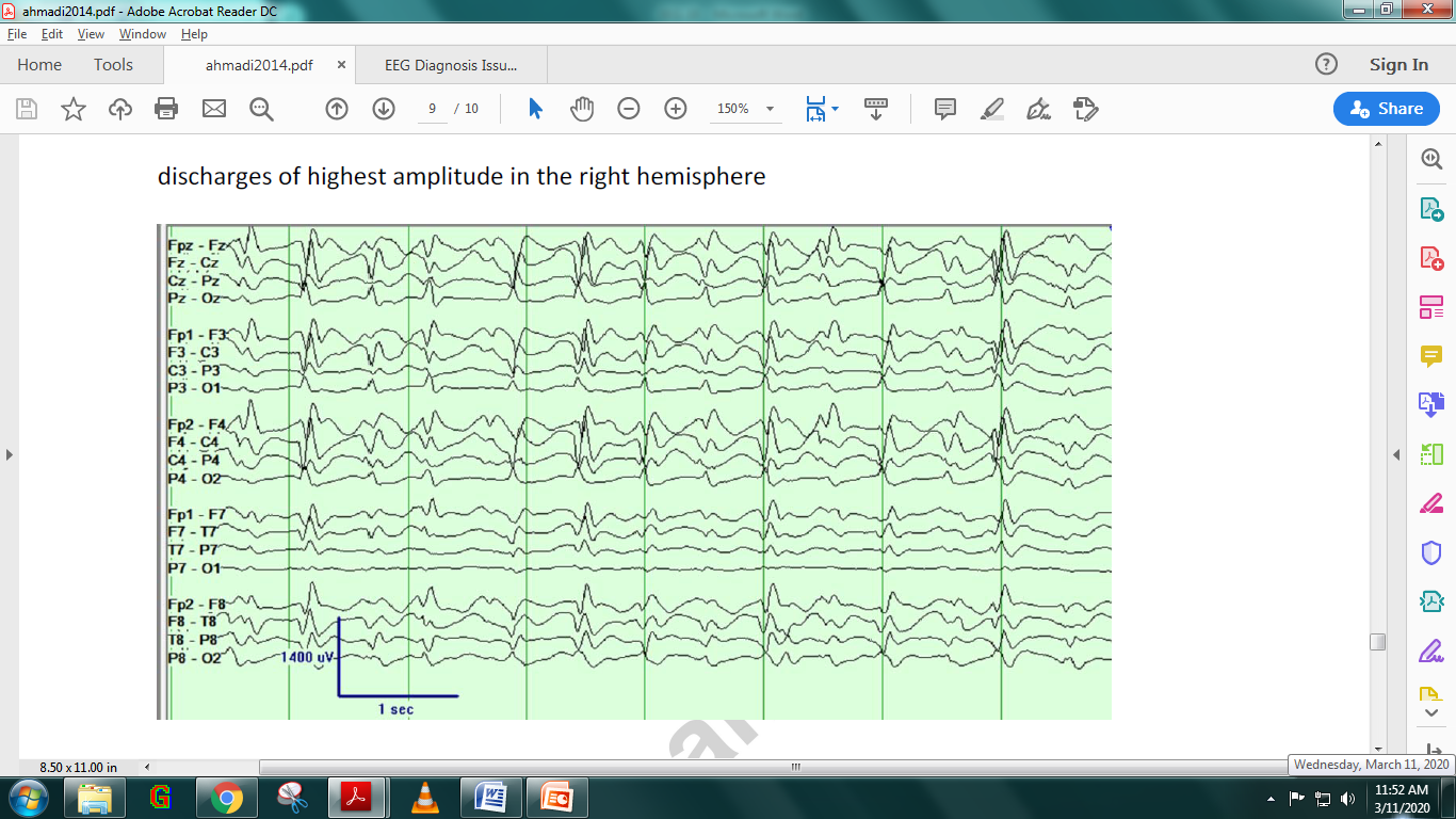
His interictal EEG showed multifocal and generalized spike wave, maximal over the right centrotemporal
region, and in sleep these discharges were nearly continuous (Figure 1).
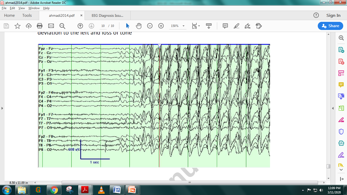
Multiple seizures were recorded which often began with right centrotemporal region theta activity but
rapidly became generalized (Figure 2).
Figure 1: Sleep EEG demonstrating electrical status epilepticus in sleep (ESES) pattern with
discharge of highest amplitude in the right hemisphere Figure 2: Seizure beginning in right centrotemporal region. During this event, the child eye
deviation to the left and loss of tone.
Diagnosis
Intractable Epilepsy induced by right hemispheric malformation
Discussion
Malformations of cortical development are one of the most common pathologies reported in surgical series of
pediatric epilepsy and frequently result in medically intractable epilepsy. They may be generalized and
diffuse, hemispheric, multifocal or focal. Severe epileptic encephalopathy and profound, early‐onset,
developmental delay is characteristic with diffuse lesions, whereas more focal lesions may present with
medically intractable epilepsy in otherwise well children and adults.
MRI advances have been instrumental in improved detection of focal cortical dysplasias, with reported
findings of increased cortical thickness, blurring of the cortical‐white matter junction, increased T2
signal, transmantle sign or localized brain atrophy. However, detection in young children prior to age 2‐3
years can be challenging due to immature myelination. The impact of intractable epilepsy in children is
significant and extends well beyond uncontrolled seizures to include intellectual disability, psychiatric
and behavioral problems and seizure related injury and death. Surgery is a viable option for children with
medically intractable epilepsy. Early surgery will allow quicker control of seizures, and minimize
associated comorbidities, thus improving long term outcome. In children with unilateral brain lesions,
seizure freedom, as well as resolution of ESES, has been shown following epilepsy surgery.
EEG Tips:
Generalized epileptiform activity on EEG may be seen in children with early onset brain lesions, and
should not dissuade from surgical intervention.
2. Focal Cortical Dysplasia in Cingulate Gyrus Linked to Seizures
Case presentation
A 25-year-old right-handed woman was presented to the clinic with an aura of sudden extreme fear before
loss of consciousness accompanied by screaming, ictal pouting and hypermotor manifestations.
The condition lasts from a few seconds to a minute.
She had her first seizure (complex partial seizures) when she was six years of age.
Investigations:
Results of neurological clinical and neuropsychological examinations were normal.
Repeated MRI revealed focal cortical thickening in the left anterior cingulate gyrus (ACG), where
glucose hypometabolism was demonstrated by PET.
Magnetoencephalography (MEG) studies revealed a cluster of abnormal interictal activity in the left
medial and lateral frontal regions.
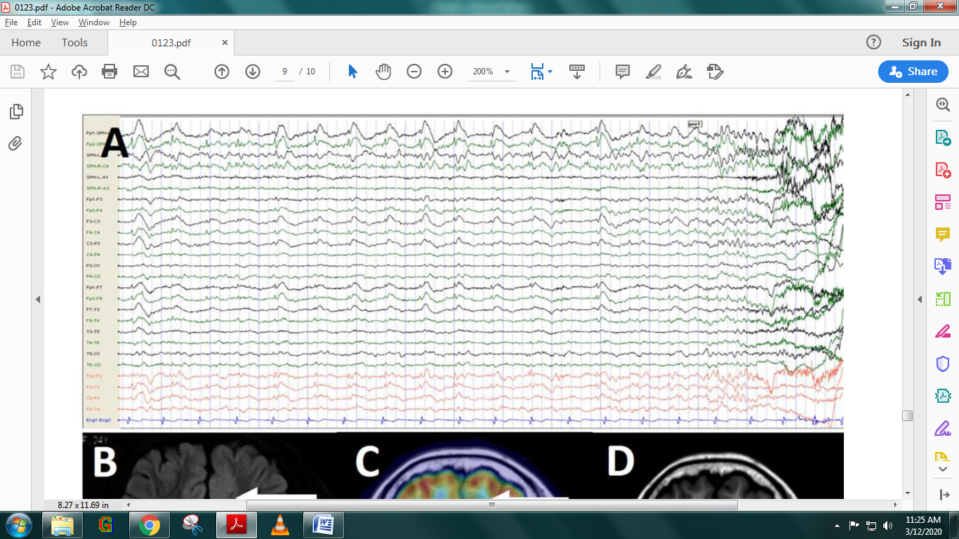
A routine scalp EEG showed symmetrical 1-2 Hz interictal epileptiform discharges and ictal low amplitude
high frequency recruiting in bilateral frontal areas (Fig. 1).
The interictal recordings showed a peculiar electrographic pattern of slow waves in the contacts of
electrode 4 implanted to the left ACG.
Multiple seizures were recorded with focal onset in the left medial frontal cortex, followed by a fast
spread to bilateral insular contacts.
Further high frequency oscillation (HFO) studies indicated that most ripple or fast ripple activities
were observed in the left medial frontal contacts of electrode 4 located at the left ACG.
High frequency (50 Hz, 0.2 ms), low intensity (0.7-2 mA) stimulation in contacts 3-8 of electrode 4
induced an aura of fear and hypermotor seizure.
Figure 1: Conventional routine EEG (electrodes placed according to the 10-20 international
system, in addition to inferior temporal electrodes) showing 1-2 Hz interictal symmetrical epileptiform
discharges and ictal low amplitude-high frequency recruiting in bilateral frontal areas.
Diagnosis
Refractory epilepsy with aura of extreme fear and hypermotor seizures
Discussion
Epilepsy, a chronic neurological disorder characterized by recurrent unprovoked seizures with varying
semiology provides a unique and valuable approach to study brain functions. In particular, localization of
seizure onset clinically manifested as extreme fear and subsequent resection of epileptogenic zone (EZ)
could help investigate the anatomical and electrophysiological mechanisms of fear production and its
spreading pattern in human brain. Temporal lobe structures, such as the amygdala, hippocampus and
parahippocampal gyrus, have been most commonly related to ictal fear in epilepsies.
However, epilepsies involving ictal fear and panic often present a technical challenge in EZ identification
because of usually poor localizing value of scalp electroencephalography (EEG), negative magnetic resonance
imaging (MRI) findings in many cases of mild focal cortical dysplasia (FCD), and complicated seizure
semiology. New imaging techniques including MEG and coregistration of MRI/PET could assist in determining
the seizure onset. Because interictal and ictal discharges of ACG, located in the middle of the brain,
propagate to both hemispheres over similar distances and thus often present bilaterally symmetrical
findings, scalp EEG might fail to reveal significant time interval between activations of the two sides.
Invasive EEG, particularly combined with HFO analysis, would be helpful in lateralizing seizure onset.
EEG Tips:
Accurate identification of EZ guided by combined analysis of HFO and PET/MRI fusion imaging, could
achieve a good clinical outcome.
3. Prolonged Refractory Status Epilepticus with Non-specific Febrile Illness
Case presentation:
A 9-year-old female presented the emergency center with stomach pain.
The patient was initially noted to be afebrile but was later found to have a fever.
She was unresponsive upon arrival.
There were no previous health problems and no family history of seizures.
Approximately 1 week prior to admission, the patient complained of “not feeling well” and had
intermittent fevers; she was also noted to be somnolent.
Investigations
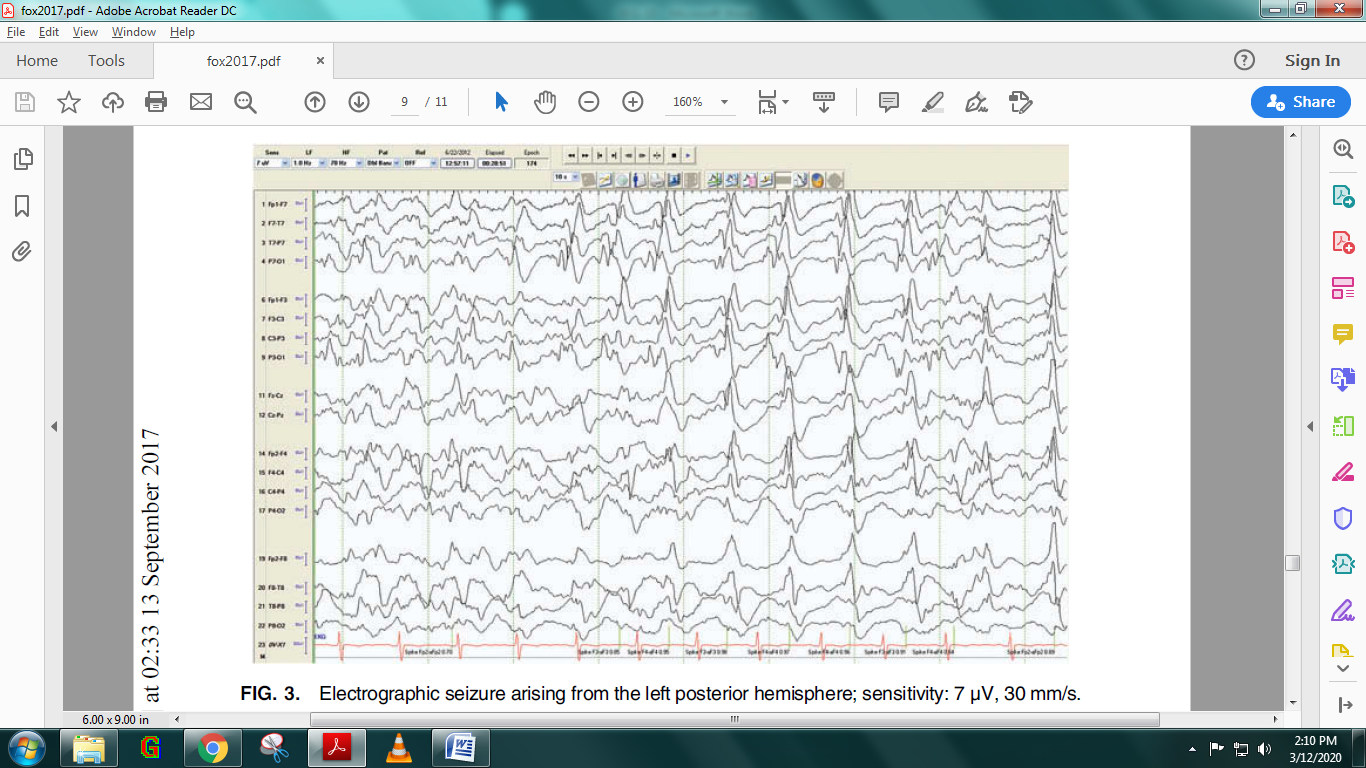
EEG revealed that the patient was having >50 seizures per day, which were left focal onset or
indeterminate onset (Figure 1).
Once it was determined that the seizures were refractory, the patient was titrated to burst suppression
with pentobarbital.
Several attempts at removing the patient from pentobarbital administration were unsuccessful.
During burst suppression, it was noted that the patient had tactile-induced seizures.
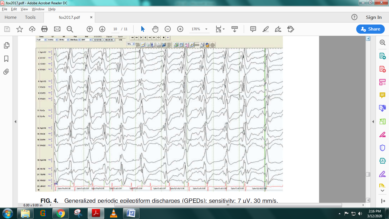
Burst suppression was weaned completely after 28 days, at which point the EEG revealed generalized
periodic epileptiform discharges (GPEDs), and no electrographic seizures were noted during the weaning
period (Figure 2).
There was a seizure-free period of 8 days before the electrographic seizures returned, originating
mostly from the left temporal region and occasionally from the right with EEG recording 2–31 seizures
per day.
Seizures continued with variable frequency until week 12 when the vEEG was removed until week 19.
Observed seizures during this period were treated with Ativan.
On week 19, an increase in seizure activity was noted and the patient was placed back on vEEG monitoring
for 10 days.
During the first 6 days, there were 7–31 subclinical seizures with right-sided predominance and
occasional secondary generalization.
After day 6, left periodic lateralized epileptiform discharges (PLEDs) were seen.
The final vEEG recording ended 21 weeks after the onset of seizure activity.
Figure 1: Electrographic seizure arising from the left posterior hemisphere; sensitivity: 7 μV,
30 mm/s.Figure 2: Generalized periodic epileptiform discharges (GPEDs); sensitivity: 7 μV, 30 mm/s.
Diagnosis
Febrile infection-related epilepsy syndrome and refractory status epilepticus
Discussion
Febrile infection-related epilepsy syndrome (FIRES) is a rare, acute, and potentially fatal childhood
epileptic syndrome. FIRES typically affects children aged 3–15 years, with a higher incidence in males. The
older age of onset differentiates FIRES from Dravet syndrome, which is also a febrile epilepsy syndrome but
has its onset during the first year of life and does not typically progress to refractory status
epilepticus. Patients present with seizures following an unremarkable febrile illness that has often cleared
1–14 days prior to the onset of seizure activity. Seizures begin and quickly develop into status epilepticus
or become very frequent, ranging from dozens to hundreds of seizures per day with loss of consciousness. The
etiology of FIRES is unknown, but there are some common clinical symptoms, such as the patient presenting
after a nonspecific febrile illness, which often clears 1–14 days prior to the onset of seizures. EEG
findings suggest that FIRES is a focal process with focal onset seizures. Ictally, EEG activity commonly
begins temporally and spreads hemispherically and/or bilaterally. No fully effective treatment for FIRES is
available, except for the use of a ketogenic diet for a few cases. The ketogenic diet can be considered at
any point, including in the early phases of FIRES.
EEG Tips:
Interictally, patients may have generalized slowing that may be considered an encephalopathic pattern
4. Intractable Epilepsy Related to Mutation in X-linked Genes
Case presentation:
A 10 year old girl was rushed to the emergency department with chief complaint of severe general
hypotonia, loss of head control, and stereotypic mouthing of both hands.
She was born to unrelated healthy parents with normal family histories.
When she was 6 months old, she developed epilepsy with tonic and tonic–clonic seizures.
Investigations:
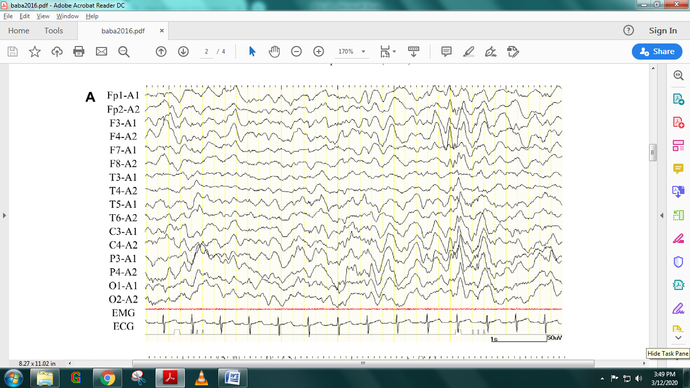
The electroencephalography (EEG) showed continuous high-voltage slow waves over the central–occipital
regions, but few epileptic discharges were identifiable (Figure 1).
Brain magnetic resonance imaging showed no structural anomalies except slight brain atrophy.
She was diagnosed with epileptic encephalopathy caused by the CDKL5 mutation.
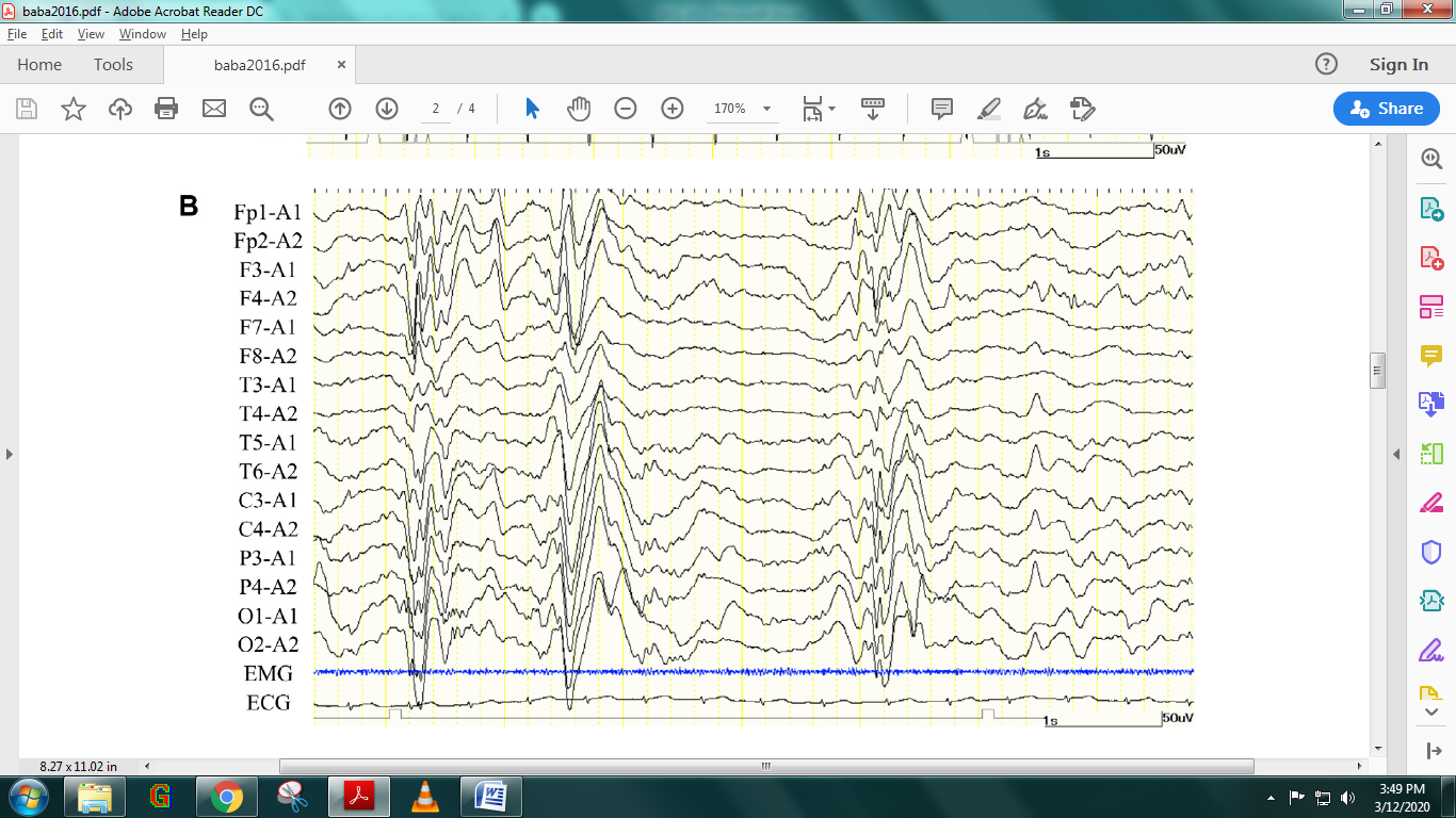
In addition to central–occipital-dominant background slowing, her interictal EEG developed paroxysmal
high-voltage positive slow waves occasionally followed by low-amplitude b activities, which were
estimated to be subclinical seizures (Figure 2).
Figure 1: EEG at 7 months showed central–occipital dominant background slowing. Figure 2: On EEG at 5 years, immediately before VNS implantation, paroxysmal high-voltage slow
waves were observed. There were spikes and waves over the central–occipital region of the right
hemisphere.
Diagnosis
Epileptic encephalopathy caused by the CDKL5 mutation
Discussion
Mutations of the X-linked gene cyclin-dependent kinase-like 5 (CDKL5) are known to cause early-onset
epileptic encephalopathy with severe neurological impairment. It is known that some patients with CDKL5
mutation experience spontaneous seizure remission with age. This condition mainly occurs in females and
often shares features of atypical Rett syndrome. In some patients, the epilepsy becomes intractable and is
resistant to conventional anticonvulsants and corticosteroid therapy. From a clinical viewpoint, the
establishment of a better management for this type of epilepsy is an urgent issue. Vagus nerve stimulation
(VNS) can be an effective adjunctive therapy for patients with drug-resistant epilepsy. In recent years,
there appears to have been more opportunities for patients with epilepsy associated with CDKL5 mutations to
receive VNS.
EEG Tips:
High-voltage positive slow waves on interictal EEGs provide some assistance for those managing cases of
CDKL5-related epilepsy.