Case 1: Extreme delta brushes in anti NMDA receptor encephalitis – muscle artefact or an EEG
phenomenon?
Case presentation
A 42-year-old woman having a history of sleep apnea syndrome presented with confusion, hallucinations,
cognitive impairment and facial dyskinesias combined with fever, headache, and diarrhea and vomiting.
A clinically visible movement pattern consisted of left hand jerks, repetitive chin movements, pursing
of the lips and blinking.
Presented here a patient with proven anti-NMDA-R encephalitis with long-term EEG monitoring.
The development of generalized seizures required intubation and temporary sedation.
Glasgow Coma Scale (GCS) score was E1M5Vtube and stereotypical movements of the hands were seen.
The subsequent course of disease included identification of an abdominal teratoma, removal of both
adnexes on day 40.
In addition, treatment with plasmapheresis (day 43-46), rituximab (4 weeks 375mg/m2 weekly) and
cyclophosphamide (6 months 750mg/m2 every month) (from day 48).
Clinical improvement became evident around day 55 (E4M6V4) with initial persistence of hallucinations
and paranoia.
Treatment with 1000 mg/day methylprednisone (day 13-17), and intravenous immunoglobins 0.4 g/kg/day (day
20-24) the rhythmic delta activity was still persisted till day 27.
As the rhythmic frontal delta activity persisted, thiopental (max 9 mg/kg) was given inducing a coma
with burst suppression EEG pattern for 48 hours (day 35-37). After this period, the rhythmic delta
activity reappeared.
6 months of cognitive rehabilitation showed improvement and enabled the patient to return home.
Investigations
Positive anti-NMDA-R antibodies in serum and CSF confirmed the diagnosis of auto-immune encephalitis.
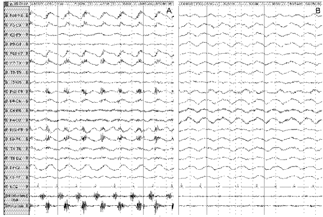
Additional electromyographic (EMG) co-registration demonstrated that extreme delta brushes resulted from
superposition of frontalis muscle EMG signal on rhythmic delta activity.
MRI showed FLAIR hyper-intensity in the insular and mesiotemporal cortex, bilaterally, and in the
anterior cingulate.
The first EEG registration (day 13) showed diffuse rhythmic frontal delta activity (encephalopathic
pattern).
EEG with video and surface EMG co-registration of the orbicularis oculi and frontalis muscles during an
episode of orofacial movements showed a delta brush pattern (Fig. 1A).
From the combined video and EMG recordings we concluded that the brush component of the delta brush most
likely represented frontalis muscle activity during the orofacial movements. Moreover, although
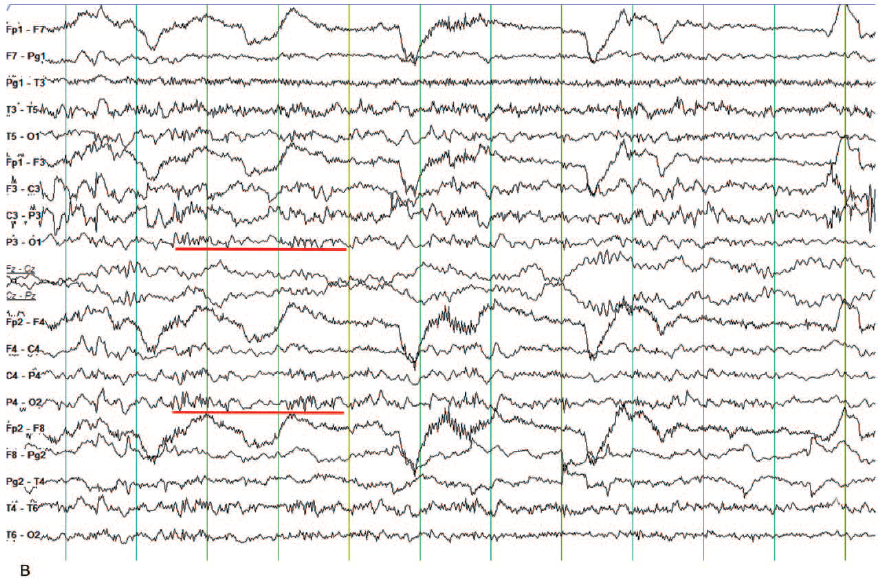
neuromuscular blocking agents were not administered, the occurrence of delta activity without brushes in
the circumstance without rhythmic frontalis muscle EMG activity strongly supports the muscular origin of
the brushes (Fig. 1B).
Figure1. EEG with video and orbicularis oculi and frontalis muscle EMG coregistration during a
five second episode of orofacial dyskinesias showing a delta brush pattern (A). During a five second
episode without EMG activity, the delta brushes are absent (B).
Diagnosis
The positive anti-NMDA-R antibodies in serum and CSF confirmed the diagnosis auto-immune encephalitis.
Discussion
This case report demonstrated EEG unique phenomenon of extreme delta brushes in anti NMDR-R encephalitis.
Here, it is proposed that the “brush” may alternatively represent an EMG artifact because of rhythmic
contractions of the frontalis muscle, occurring in synchrony with frontal delta activity. The alternating
contractions of the frontalis and orbicularis oculi muscles, synchronously with the EEG delta activity,
suggest that both phenomena may arise from a common generator mechanism, probably involving subcortical
structures. The frequent presence of oro-lingual-facial dyskinesias in anti-NMDA-R encephalitis which may
occur both clinically and subclinically as demonstrated in this patient.
The rhythmic frontalis muscle contractions were not always clinically visible in this patient, here it is
emphasized that for assigning extreme delta brushes as an EEG pattern, EMG co-registration is required to
rule out an EMG ‘artifact’. Studies with EEG-EMG co-registration and administration of a neuromuscular
blocking agent are useful to investigate the nature of extreme delta brushes
Case 2: Encephalitis in a traveller with typhoid fever: efficacy of corticosteroids
Case presentation
A 19-year-old French man without medical history was admitted for fever after a 2-week backpacking trip
in the Philippines with his parents and sister.
He was a student, living in France with his parents and did not report use of alcohol, tobacco or any
other drug.
After his return, fever up to 40°C had started without gastrointestinal symptoms and abdominal signs for
10 days.
Before admission, after the self-treatment with paracetamol and a non-steroidal anti-inflammatory
regimen for 10 days the fever was still persisted.
On Day 1, clinical examination was normal without any neurological sign but showed pyramidal irritation
with brisk, diffuse, polykinetic reflexes of the lower limbs, a plantar reflex in triple flexion and
positive Hoffman’s test of the upper limbs.
On Day 2, blood cultures isolated Salmonella enteric serotype Typhi and treated with intravenous
antibiotic therapy.
On Day 4, patient was afebrile switched to oral ofloxacin 200 mg twice a day. At Day 5, the patient was
discharged.
On Day 7, reappearance of fever (38.5°C) led to new admission. On Day 8, his neurological status got
worse, with appearance of pyramidal signs of the upper limb, and disturbances in language functions,
agitation then confusion.
On Day 9, patient started his usual activities again. On Day 11, he walked alone, was coherent and able
to read books again and discharged.
Clinical investigations:
Electroencephalogram (EEG) signal showed nonspecific signs of cortical hyper excitability with sharp
slow waves and slow waves mainly in the frontal regions compatible with anencephalopathy (Figure 1).
Cerebral magnetic resonance imaging (MRI) did not show abnormalities and excluded acute demyelinating
encephalomyelitis (ADEM) whereas CSF culture excluded bacterial meningo-encephalitis.
Moreover, blood sample analysis showed negative for stool culture and for Dengue and malarial fever.
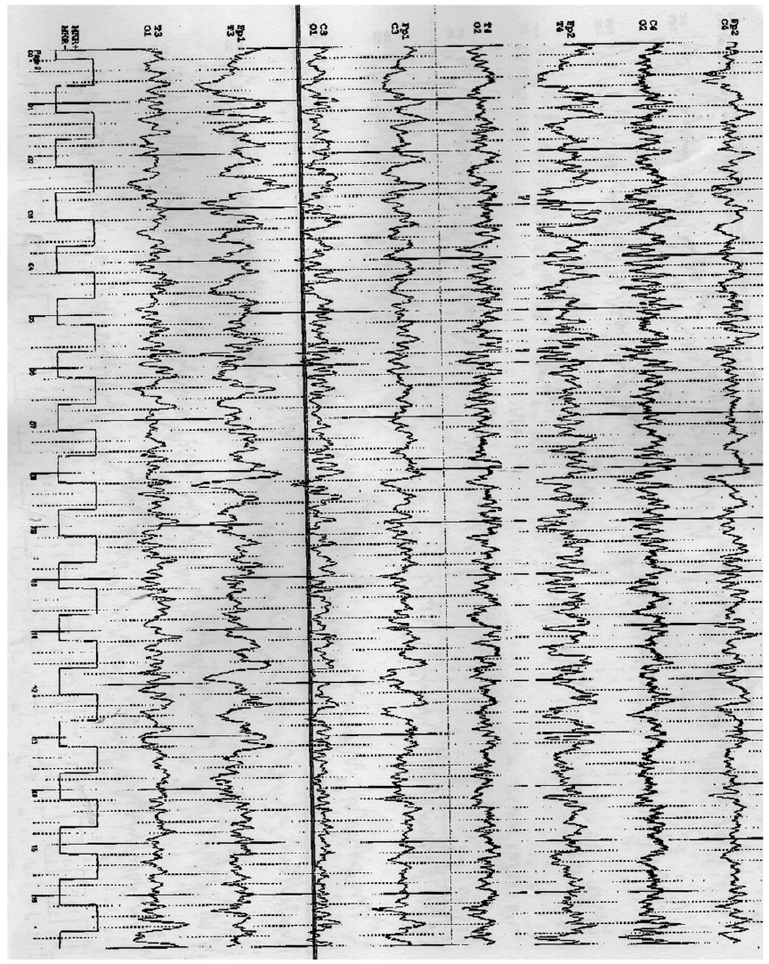
Electroencephalogram of the traveler patient
Diagnosis
EEG confirmed the diffuse encephalitis in the traveler with typhoid fever.
Discussion
This case, as evidenced by EEG results, highlights the need to understand pathophysiology and management of
this diffuse encephalitis. Because, clinical management is often difficult as Western physicians are not
used to diagnose this infection or treat such patients. According to the authors, this is the first case
where EEG signs are described which is understandable given its poor availability in developing countries.
The traveller gets encephalitis after treatment with antibiotics for 4 days raising the possible role of
typhoid endotoxemia.
Covering a wide scope of CSF results reported in the literature in cases of encephalopathy complicating
typhoid fever. As in the present case, CSF may be abnormal. On the other hand, cases have been reported with
a normal MRI brain as in this case. Cerebellar oedema and venous congestion of brain cells are often evident
in typhoid fever’s encephalopathy. The initial response was good with ceftriaxone and the antibiotic
sensitive Salmonella makes antibiotic failure unlikely. In addition, the neurological status of the patient
only improved after corticosteroid treatment had been instituted. Such reactions are considered as a direct
result of the damaging effect of the toxins released by S. enteric (whichever serotype is Typhi or
Paratyphi) during bacterial lysis with typhoid toxins having an important affinity for the central nervous
system.
In cases of severe typhoid fever, high doses of corticosteroids like dexamethasone should be considered to
decrease the mortality rate. Finally, early recognition and appropriate management of complications
associated with typhoid fever should reduce morbidity and mortality, especially for travellers returning
from endemic areas.
EEG Tips:
Electroencephalogram (EEG) signal revealed nonspecific signs of cortical hyper excitability with sharp
slow waves and slow waves in the frontal regions compatible with anencephalopathy.
Reported as the first case where these EEG signs are described which is understandable given its poor
availability in developing countries.
Case3: Neurosyphilis mimicking autoimmune encephalitis in a 52-year-old man
Case study:
A 52-year-old man in a long-term sexual relationship with the same gender presented here with agitation,
confusion and difficulty in speaking for about two weeks.
On evaluation, his vital organs were found to be normal with agitation and global aphasia.
On a screening neurologic examination, no focal deficits were observed.
On phenytoin treatment, seizure activity was ceased but with the persistence of same symptoms of
agitation, confusion and aphasia.
Six months before he was admitted with the symptoms mentioned above with an episode of vertigo.
Case Investigations
Initial electroencephalogram showed left posterior temporal slowing and without any seizure activity.
Lumbar puncture showed inflammation in cerebrospinal fluid (CSF) infiltrated with leukocytes and
lymphocytes, with the elevation in protein levels.
CSF bacterial culture, polymerase chain reaction of herpes simplex virus, serum HIV tests were all
showed negative results.
Repeated EEG showed intermittent seizure activity after lacosamide 200 mg and levetiracetam 500 mg given
orally twice a day.
Repeated brain MRI showed left hippocampal atrophy without any abnormality.
Repeat serum anti-glutamic acid decarboxylase(GAD) antibody was again positive, and a repeat lumbar
puncture yielded results similar to those described above. No empiric antimicrobial therapy was
administered.
A diagnosis of autoimmune encephalitis was considered after subacute neurologic decline with seizures,
medial temporal lobe signal abnormality on initial MRI, and positive serum anti-GAD antibody.
Serum analysis confirmed neurosyphilis by using treponemal automated enzyme immunoassay, Treponema
pallidum particle agglutination assay, quantitative rapid plasma reagin values, and CSF quantitative
venereal disease research laboratory test (VDRL).
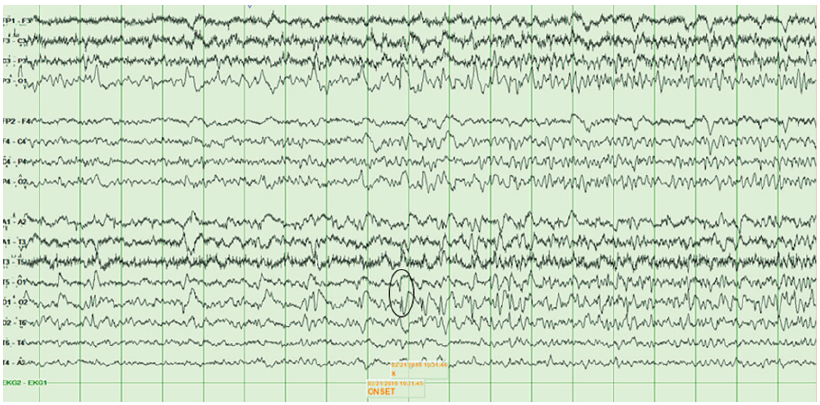
EEG showing the onset of left temporal-occipital seizure in neurosyphilis at T5-O1 and O1-O2
(circle),and propagation entirely in the left temporal lobe.
Diagnosis
Positivity of serum anti-GAD antibody with seizures and medial temporal lobe abnormality in MRI confirmed
autoimmune encephalitis
Discussion
Here presented an unusual case of neurosyphilis mimicking rare autoimmune encephalitis. Autoimmune
encephalitis is the second most common cause of encephalitis with temporal lobe abnormalities. Therefore, it
is a major diagnostic consideration in patients like in this study.
Clinicians should keep consider that intrathecal antibody production may also be seen with numerous central
nervous system infections like neurosyphilis. Although neurosyphilis frequently occurs in HIV-infected
persons, patients with neurosyphilis mimicking autoimmune encephalitis are typically not infected with HIV.
The interface of infectious and autoimmune encephalitis has reached the forefront of neurology in recent
years, as evidenced by the discovery of anti-NMDAR encephalitis triggered by herpes simplex virus
encephalitis. Success of antimicrobial therapy is marked by clinical improvement or stabilization, decline
in CSF leukocyte count at six months and resolution of all CSF abnormalities at two years. Future study in
this field may elucidate complex mechanisms of disease in patients such as this case.
EEG Tips:
EEG showed onset of left temporal-occipital seizure in neurosyphilis.
EEG showed left posterior temporal slowing without seizure activity.
Case 4: Occipital epilepsy versus progressive myoclonic epilepsy in a patient with continuous
occipital spikes and photosensitivity in electroencephalogram
Case study:
A 21 years old female patient with refractory epilepsy seizures for 7 years is presented here.
At the age of 14, this patient has a tonic-clonic seizure with the frequency of 3-4 times in a year and
the frequency increased drastically up to 12 times in a year.
Simultaneously, the patient exhibited serious language problems, with difficulty in balancing and
walking.
Because of her poor development she has dropped-out at the age of 16 from the school despite her
babyhood being normal.
Case investigations:
EEG and magnetic resonance imaging (MRI) were performed on this patient.
MRI confirmed diffused atrophy in cerebral cortex in the frontal and parietal lobes.
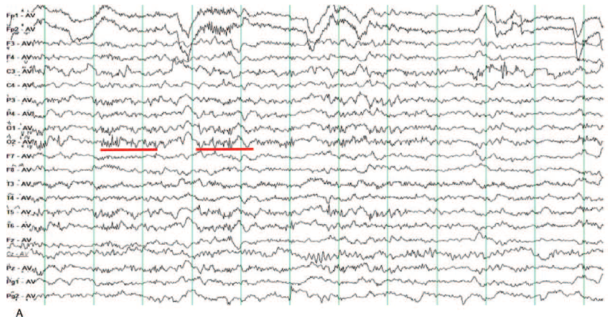
EEG displayed slower activity in background with continuous spike in occipital lobe.
Ictal EEG conveyed generalized polyspike and spike-slow complex wave synchronized to the tonic-clonic
seizure.
EEG changes showed slow background with generalized polyspike and spike-slow complex discharges
indicated severe epileptic syndrome or epileptic encephalopathy.
Figure 1A. Slow activity in background with continuous spike or spike-slow wave in occipital
lobe Figure 1B. Continuous spike or spike-slow wave presented in bipolar montage
Diagnosis
Photosensitivity in EEG combined with the voice-sensitive or light-sensitive seizure onset was helpful in the
diagnosis of PME.
Discussion
Progressive myoclonic epilepsy (PME) is a complex, rare disorder or syndrome caused by metabolic, genetic,
and neurodegenerative diseases, characterized by refractory epilepsy, neurological deterioration, and
cognitive impairment with a poor prognosis. Using special EEG manifestations mentioned in case
investigation, a girl patient diagnosed as PME syndrome. It recurrently causes diagnostic problems with its
extensive diagnostic entities. In occipital epilepsy, It is very common to have posterior discharges of
spike or spike-slow, however, persistent low-amplitude poly-spike discharges were not usual morphology.
Similar EEG changes described in this case study offer valuable guidance to clinicians and make it easier to
limit the differential diagnosis.
EEG Tips:
This case showed special EEG findings in PME, which may be confused with occipital epilepsy or
photosensitive epilepsy.
Helps clinical doctors to use EEG in establishing the suspected diagnosis of PME as soon as possible
Photosensitivity in EEG combined with the voice-sensitive or light-sensitive seizure onset was also
helpful in the diagnosis of PME.