1. Right orbitofrontal epilepsy featuring ictal swearing
Case presentation
A 36-year-old, right-handed man was referred for pharmacoresistant epilepsy with seizures occurring every night.
His seizures started at the age of 26 years old, refractory to several antiepileptic drug regimes. His seizures attacked with no aura, the majority of seizures were characterized by sudden awakening and quickly followed by swearing and repetitive purposeless movements (right upper limb).
There was no history of perinatal complications, febrile convulsion, CNS infection or trauma.
Family history was also uneventful.
Investigations
Scalp video-EEG (VEEG) monitoring revealed the interictal spikes were located in the left temporal and frontal region (SPH-L, F7, T3, F3), the spikes appeared not frequently.
After five days of VEEG, eight stereotypical seizures were recorded, one of which evolved into a generalized tonic-clonic seizure.
Ictal EEG consistently showed low amplitude theta frequency activity in right temporal region followed by diffuse spike–wave discharges.
Intracranial evaluation was performed using stereotactic electroencephalography (SEEG). Ten depth electrodes were implanted to sample targets in bilateral hemispheres. Three electrodes were implanted in left insular cortex, anterior cingulated cortex and orbitofrontal cortex. Three electrodes were implanted in the left temporal lobe and hippocampus.
In the right hemisphere, four electrodes were implanted in the posterior part of the right gyrus rectus, anterior cingulate cortex, insular cortex, temporal cortex and hippocampus.
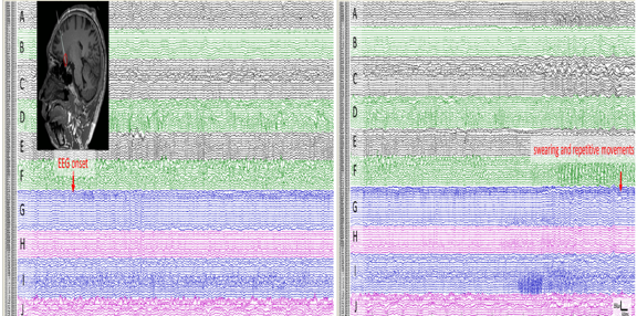
Intracranial EEG monitoring showed interictal spikes were located in the left temporal lobe and right gyrus rectus. EEG onsets were characterized by paroxysmal fast activity arising from the right gyrus rectus, followed by rhythmic activities in the right dorsolateral prefrontal cortex and left temporal region.
There were eleven seizures were recorded via SEEG, ten of them (90.9%) featured ictal swearing, the mean delay from EEG onset to appearance of swearing was 25.4s.
When the discharge involved the right dorsolateral frontal cortex and the left temporal region, the swearing and repetitive limb movements appeared. Cortical stimulation (0.2 ms width pulse, 50 Hz, 1–6 mA, 3 s duration) of electrode contacts in the EEG onset region did not elicit swearing but repetitive purposeless movements in right upper limb which was consist with stereotypical seizure.
The patient underwent a resection of the anterior part of right prefrontal lobe including the frontal pole, gyrus rectus and subgenual cingulate cortex.
Histopathology demonstrated a focal cortical dysplasia (FCDIIb)
The patient has been seizure free for 8 months at last follow up.
Figure 1. SEEG of an ictus. (A): Ictal low voltage fast activities originate from the right gyrus rectus recorded by the electrode G (G1-G5, arrow). (B): When the discharge involves the right dorsolateral frontal and left temporal regions, the swearing and repetitive limb movements appears (arrow). The delay from EEG onset to appearance of swearing is about 20s.
Diagnosis
Ictal swearing and repetitive motor automatisms due to non-dominant right frontal lobe epilepsy
Discussion
This case report describes the first surgically proven report of ictal swearing originated from right gyrus rectus. SEEG electrodes were implanted in the bilateral frontal, temporal and insular lobes, the result demonstrated that the EEG ictal onset was localized in the right gyrus rectus, when the seizure activity involved the right dorsolateral prefrontal cortex and left temporal region, the ictal swearing appeared. Ictal swearing is probably related to the recruitment of an orbitofrontal–temporal networks activated by the epileptogenic zone. The favorable post-surgical outcome is further proof of seizure onset zone localization.
It is proposed that swearing is a form of emotional or compulsive speech, and there were two possible mechanisms underlying ictal speech automatisms: 1. ictal speech results from release of the dominant hemisphere from the inhibitory action of the non-dominant one; 2. ictal speech results directly from epileptic activity of the non-dominant hemisphere. Ictal swearing is a kind of verbal automatism correlated with negative emotional experience; it has been known that the ventral system, including the amygdala and ventrolateral prefrontal cortex, are associated with negative emotional processing. It has been also proposed that an inhibitory connection from the orbitofrontal cortex to the amygdala regulates negative emotion and related behaviors.
In this case, the triggered seizures during cortical stimulation manifested only right upper limb repetitive purposeless movements without swearing. It is an additional proof that ictal speech manifestation is probably produced by the recruitment of broad and complex neural networks.
EEG TIP
Orbitofrontal–temporal network is closely associated with symptomatology of ictal swearing.
REFERENCE: Zhang X, Zhang G, Ma K, et al. A case of right orbitofrontal epilepsy featuring ictal swearing. J Neurol Sci. 2019 Feb 15;397:1-3.
2. Childhood absence epilepsy with focal spikes
Case presentation
A 6-year-old right-handed male presented to the clinical setup on March 2011 due to frequent paroxysmal daze. The main behavioral manifestations were daze, absence and no response to calling.
Each attack would last for approximately 10 seconds, with more than 10 of such episodes each day during the waking state, without falling or convulsions. The patient was born at full term and exhibited normal physical and mental development.
No family history of absence seizures was noted.
No genetic evidence was available, although the parents were not close relatives.
In January 2011, the patient started oral sodium valproate sustained-release tablets at 250 mg/day, and the dose was gradually increased to 750 mg/day.
Despite the dose increase, the patient continued to suffer from 4−5 attacks per day.
In March of the same year, lamotrigine was started. The dose was gradually increased to 25 mg/day, and the attacks completely disappeared.
Furthermore, the sodium valproate dose was gradually reduced and subsequently withdrawn, while the dose of lamotrigine was gradually increased to 50 mg/day.
Lamotrigine treatment was continued with no further reports of seizure episodes or adverse effects.
Investigations
The physical and neurological examination results were all normal.
The brain MRI was also normal.
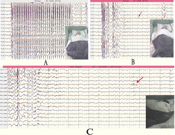
The 24-hour video EEG examination(Fig. 1A) revealed frequent 2.5−3.0 Hz generalized spike-and-wave complexes that lasted for 10−15 seconds, with sporadic spikes also observed at the left central area (Fig. 1B-1C).
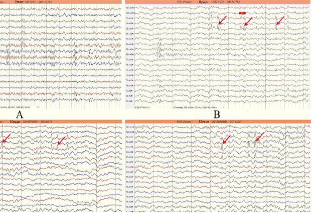
The EEG examination in July 2011(Fig. 2A) demonstrated that the 3-Hz generalized spike-and-wave complexes disappeared in the waking state.
Repeated blood tests revealed no abnormalities in liver function.
The video EEG examination in July 2012 (Fig. 2B) revealed frequent spike-waves in the left central area during sleep, but no widespread abnormal discharges occurred when the patient was awake.
During this period, no seizures occurred, and the patient demonstrated normal sleep quality and mental development, with excellent academic results.
Lamotrigine medication was continued until 2014, and the repeated video EEG revealed spikes mainly at the left central area during sleep (Fig. 2C).
At that time, no attacks occurred for nearly three years. Hence, the lamotrigine dose was gradually reduced with final withdrawal in 2015.
The repeated video EEG examinations in 2016 continued to reveal spikes at the left central area, but these disappeared in 2017, at which time medication had been withdrawn for 2.5 years with no subsequent seizure attacks.
Figure 1. Results of the first video EEG showing concomitant synchronous spike-and-wave complexes plus left focal spikesFigure 2. Post-treatment video EEG results showing residual spikes
Childhood absence epilepsy is a common generalized epilepsy syndrome with onset at school age. Absence is the main behavioral manifestation, and the corresponding electroencephalogram (EEG) is characterized by bilateral, symmetrical and synchronous 2.5−4.0 Hz of spike-and-wave complexes. However, generalized spike-and-waves (3 Hz) with focal central discharges are relatively rare.
The case patient demonstrated features consistent with childhood absence epilepsy: repeated daze and absence episodes during the waking state starting at school age, attacks that lasted for approximately 10 seconds without convulsions or falling, and 2.5-3.0 Hz of generalized spike-and-wave complexes continuing for 10−15 seconds. However, the sleep-state EEG revealed concomitant abnormal discharges without seizures at the central area, which is the sensory-motor area of the hemispherical convex surface corresponding to the central area of the international 10-20 system.
At first, the investigators considered that it was a fragmented change in general discharge. However, when the generalized epileptiform discharges disappeared during the waking period after the patient's condition was completely controlled, focal spike waves were still observed in the left frontal central area in the sleep stage. Therefore, this focal spike wave could not be simply explained as the fragmentation of generalized epileptiform discharges, since it should be a coexistence of generalized epilepsy syndrome with focal discharge.
EEG TIP
At first, the investigators considered that it was a fragmented change in general discharge. However, when the generalized epileptiform discharges disappeared during the waking period after the patient's condition was completely controlled, focal spike waves were still observed in the left frontal central area in the sleep stage. Therefore, this focal spike wave could not be simply explained as the fragmentation of generalized epileptiform discharges, since it should be a coexistence of generalized epilepsy syndrome with focal discharge.
REFERNCE: Yu YL, Shi MT, Lu HJ. A case of childhood absence epilepsy with focal spikes. World Neurosurg. 2019 Mar 14. pii: S1878-8750(19)30707-7.
3. A Unique Form of Reflex Epilepsy Induced by Exiting Water
Case presentation
A 13-year-old developmentally normal boy with no identifiable epilepsy risk factors presented with a 3-year history of seizures. Seizures did not occur while the patient was immersed in water.
Approximately 20 seconds after exiting water (shower, bath-tub, or pool), he experiences an ice-cold sensation that starts in his lower spine and rises toward the bitemporal head regions. This evolves into a sensation of vertigo and loss of consciousness followed by right eye deviation, right head version, and subsequent generalized tonic-clonic activity.
His seizures are not modulated by length of water exposure, the temperature of either the water or external environment, or specific annual seasons.
Investigations
His neurological examination was normal.
His interictal electroencephalogram (EEG) revealed a normal awake background and sleep activated left frontal spike discharges.
An ictal event was captured in the epilepsy monitoring unit by showering the patient in lukewarm water, followed by the transferring him to a room set at 24°C without additional tactile stimuli such as towels.
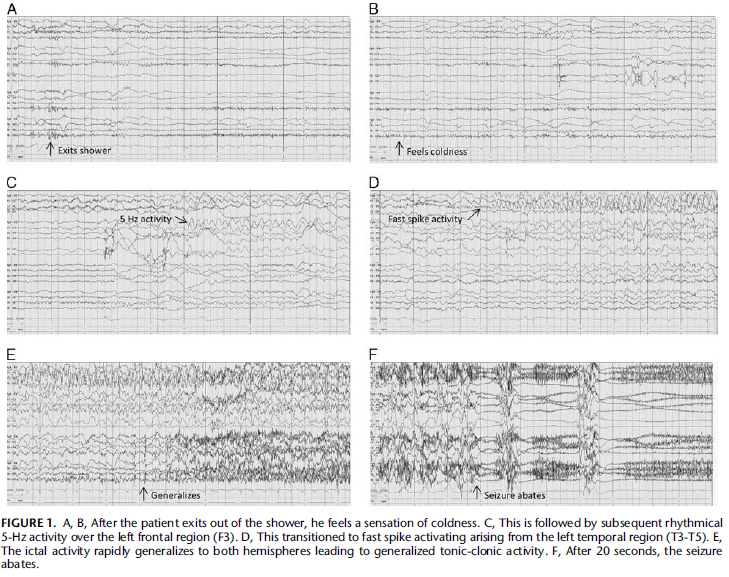
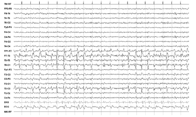
The clinical onset of a sensation of coldness preceded the electrographic onset of rhythmical left frontal 5-Hz activity that evolved into left temporal spikes, then generalizing to both hemispheres (Fig. 1).
There were no electrocardiogram abnormalities during the event.
A brain magnetic resonance image revealed normal brain parenchyma and an incidental left paramedian small arachnoid cyst.
Diagnosis
Reflex epilepsy induced by exiting water
Discussion
Reflex epilepsies represent a form of epilepsy in which unique modes of seizure precipitation are characterized by endogenous or exogenous stimuli. Hot water epilepsy represents a subtype of reflex epilepsy in which seizure precipitation arises from the act of immersing the head with hot water.
This case demonstrates exclusive reflex epilepsy. The patients had focal-onset seizures with secondary generalization. The clinical semiology initiated as a cold sensation in the body, followed by convulsive or atonic activity. In the child, the clinical onset preceded the electrographic onset, suggesting a mesial seizure focus with spread toward the left frontotemporal cortex.
It is hypothesizes that exiting water results in seizure provocation by a sudden drop in temperature, activating left somatosensory and insular territories that are involved in thermosensory processing.
The patient demonstrates a unique form of bathing epilepsy that is provoked by the removal of the body from water. This condition seems to manifest with focal-onset seizures.
EEG TIP
The presence of left temporal ictal activity corresponds with reported cases of both hot water epilepsy and bathing epilepsy.
REFERENCE: Appavu B, Fox J, Little R et al. Seizures Induced by Exiting Water: A Unique Form of Reflex Epilepsy. Neurologist. 2017 Sep;22(5):196-198.
4. Periodic EEG Pattern Associated with Ictal Manifestation
Case presentation
A 66 year-old right-handed man with no familiar history of epilepsy presented in Emergency Unit because of sudden onset of language disturbance associated with hypersalivation and paraesthesia at the right forehand about 20 min before the arrival.
The anamnestic record showed the presence of similar brief episodes in the previous days, sometimes associated with a sense of laryngeal constriction.
Investigations
During the neurological examination, the patient was restless and showed a fluent aphasia with an incongruous and meaningless speech and the inability to comprehend simple instructions.
Aphasia Rapid Test (ART) was performed resulting in total score of 23/26 points.
With exception of paraesthesia at the right forehand, there were no other sensory-motor deficits.
Cranial nerves examination was normal.
A brain Computed Tomography (CT) scan was performed that showed an hypodense lesion within the insular-temporal region of the left hemisphere.
A 21 derivations EEG recording was then performed which showed mid-temporal continuous spike-wave complexes at 1.5–2 Hz and 100–150 uV of amplitude with phase opposition on T3 with superimposed fast activity (Lateralized Periodic Discharges plus superimposed fast activity, LPDs + F pattern), (Fig. 1).
The patient was then treated with a bolus of Lorazepam 10 mg and underwent to continuous EEG monitoring. Over the next 30 min there was a complete normalization of the EEG abnormalities with a resolution of all the neurological symptoms including aphasia (ART = 0/26).
Figure 1. EEG findings during the ictal phase. Shows periodic spike-wave complexes at 1.5–2 Hz and 100–150 uV of amplitude with phase opposition on T3 mixed with polyspikes, described as Lateralized Periodic Discharges plus superimposed fast activity (LPDs + F) on the left fronto-temporal derivations.
Diagnosis
Lateralized periodic discharges in insular status epilepticus
Discussion
Language disturbances can be usually found in various pathological acute pictures involving the dominant frontal and temporal lobes. In the present report, aphasia was the main manifestation of the focal status epilepticus (SE) in association with other symptoms related to insular involvement. Routine EEG was helpful in demonstrating the epileptic origin of the symptoms though the hidden distant location of insular lobe cortex compared with the localization of the scalp electrodes.
According to recent evidences the direct stimulation of the insular lobe induces some characteristic symptoms that can appear in individuals with insular epilepsy. In particular, sense of laryngeal constriction, paraesthesia, dysarthria, auditory hallucinations, somatosensory auras, autonomic nervous system symptoms, hypermotor crisis and retrosternal pain could represent the leading signs of insular lobe epilepsy.
The present case report stresses the electro-clinical correlation between insular status epilepticus and Lateralized Periodic Discharges (LPDs). LPDs pattern is usually correlated with structural lesions of cortical or subcortical areas due to some pathological conditions such as acute stroke, brain tumours, infections, traumas and metabolic diseases. The fluctuating feature and its correlation with a suggestive EEG pattern should raise suspicion that aphasia might be an ictal manifestation rather than merely a focal sign due to a cerebral lesion.
EEG TIP
Insular lobe seizures generally represent a misconceived ictal phenomenon characterized by specific neurological signs.
Aphasia can be a rare presenting sign associated with insular lobe epilepsy which could be easily mistaken for a manifestation of other acute brain diseases.
REFERENCE: Dono F, Russo M, Carrarini C et al. Lateralized periodic discharges in insular status epilepticus: A case report of a periodic EEG pattern associated with ictal manifestation. Clin Neurophysiol Pract. 2019 Jan 24;4:27-29.