1. A 15-year-old boy with acute headache, blurred vision and a secondarily generalized clonic
seizure.
Case presentation
A 15-year-old boy with Wegener granulomatosis and chronic renal failure was started on hemodialysis and
plasma exchange.
On day 7 of dialysis, he developed a severe throbbing bifrontal headache associated with nausea,
vomiting, and transient blurred vision.
After a 3-hour period of blurred vision, he began to have focal seizures characterized by staring,
altered awareness, and repetitive jerking of the eyes to the left side lasting up to 2 minutes.
After the seizure, he was drowsy but responded to voice and was oriented; however, his attention was
poor.
His speech was fluent, and he was able to process two-step commands.
He reported profound visual loss, and he could not count fingers or see light.
His pupils were equal and reactive to light.
Investigations
His blood pressure ranged from 150/90 to 170/90 mm Hg.
No focal neurological findings were documented on examination immediately after the seizure
Fundoscopic examination was normal, and extraocular movements were full.
The remainder of the neurological examination was normal.
Computed tomography of the head was normal.
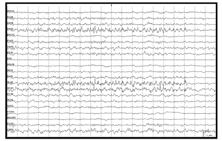
An EEG performed showed eight electrographic seizures, lasting from 18 to 128 seconds, during a
30-minute recording.
They originated independently in the right and left occipital areas with a field to the posterior
temporal-parietal areas.
During these electrographic seizures, there were no changes in patient’s behavior (Fig. 1)
FIG. 1. Ictal EEG recording in a bipolar montage (parasagittal and posterior halo) demonstrating
a right occipital seizure with an evolving rhythm with spike fast activity in the right occipital area.
MRI of the brain 48 hours after onset of symptoms showed bilateral parietal–occipital and temporal T2
hyperintense signal changes predominantly involving white matter.
There were also T2 hyperintense signal changes in the left centrum semiovale and the right frontal
subcortical region.
Diffusion-weighted images and apparent diffusion coefficient were consistent with vasogenic edema.
Posterior reversible encephalopathy syndrome (PRES) is rare in the pediatric population and is characterized
by reversible vasogenic edema in the white matter of the posterior parietal–occipital regions of the brain.
It is most commonly associated with the administration of immunosuppressive and cytotoxic agents and
malignant hypertension. The clinical manifestations are characterized by headache, nausea, vomiting,
decreased level of consciousness, seizures, and visual disturbances (cortical visual impairment,
hemianopsia, and others visual manifestations). Similar symptoms may occur in occipital lobe seizures.
Occipital lobe seizures is the major clinical manifestation of PRES. It may cause severe ictal visual loss
through a myriad of mechanisms including cytotoxic injury and vasogenic edema, leading to possible
triggering of an inflammatory cascade, membrane instability, and ultimately an imbalance of excitatory and
inhibitory neurotransmitters. These pathways could lead to excessive neuronal excitation and thus
epileptogenesis resulting in occipital lobe seizures. Ongoing seizures may contribute to further brain
injury.
Generalised slowing and inter-ictal discharges—not focal seizures or status epilepticus—are the most common
EEG findings.
The ictal video EEG recordings shows a diffuse slowing and focal unilateral temporo-occipital ictal activity.
2. A 3-month-old male infant with multiple afebrile seizures and midgut malrotation.
Case presentation
A 3-month-old male infant was referred to hospital following two episodes of generalized tonic-clonic
seizure without fever.
He was born via a normal vaginal delivery at 38 weeks and 3 days of gestation, with a birth weight of
3900 g.
The perinatal birth history was unremarkable, and there was no family history of epilepsy or febrile
seizure.
He had intermittent abdominal distention and postprandial projectile, non-bilious vomitus from birth.
Investigations
On admission, the vital signs were blood pressure of 103/76 mm Hg, pulse rate of 156 beats/min, body
temperature of 36.4 C and a respiratory rate of 28 breaths/min.
His body weight was 6 kg (25 percentile), height was 65 cm (95 percentile), and head circumference was
42 cm (90 percentile).
He was alert and there were no other abnormal neurological findings.
Blood analysis revealed 10.6 g/dl hemoglobin, 15,420 leukocytes/mm3 (49.7% neutrophils, 32.5%
lymphocytes, and 16.9% monocytes) and 583,000 platelets/mm3 .
Serum electrolyte levels were 131 mEq/l sodium, 5.0 mEq/l potassium, and 87 mEq/l chloride.
Stools were negative for rotavirus antigen, and no bacteria were identified in the blood, urine, CSF
or stools.
The CSF was negative for Herpes simplex virus according to PCR analysis, and a brain CT was normal
findings.
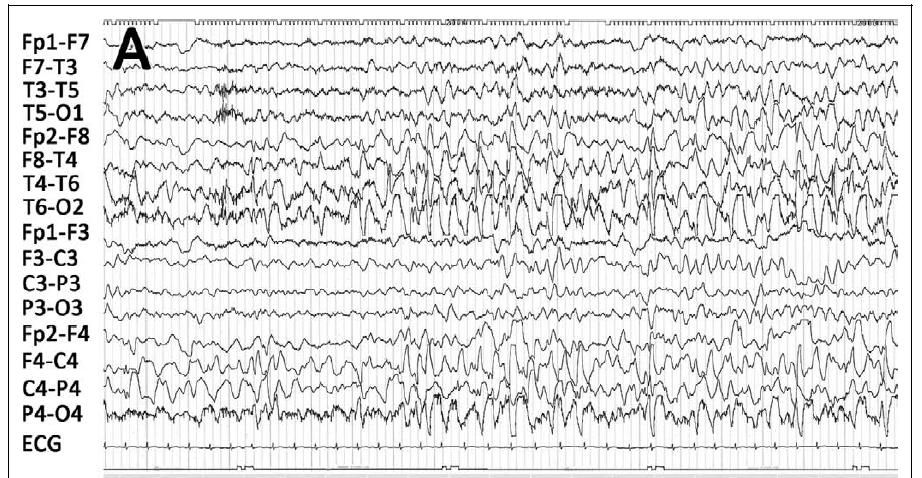
An EEG showed slow and disorganized background rhythms and frequent sharp wave discharges from both
posterior quadrants (Fig. 1). A general tonic–clonic seizure occurred several hours later.
Fig. 1. An EEG showing slow and disorganized background rhythms and frequent sharp wave
discharges from both posterior quadrants.
A simple abdominal X-ray revealed an abnormal gas pattern.
Under suspicion of a gastrointestinal problem, an abdominal ultrasonography was performed, and the
findings suggested a midgut malrotation
Diagnosis
Benign convulsion with mild gastroenteritis (CwG)
Discussion
Provoked seizures are a common pediatric problem, and can be caused by a variety of diseases. Gastroenteritis
can cause benign infantile seizures, also known as benign convulsion with mild gastroenteritis (CwG). CwG
manifests as afebrile generalized convulsions with symptoms of gastroenteritis in otherwise healthy children
aged 6 months to 3 years. CwG patients are normal in terms of blood glucose, serum electrolytes,
cerebrospinal fluid (CSF) and electroencephalogram (EEG) findings. CwG can be associated with
gastroenteritis caused by viruses such as rotavirus, norovirus, sapovirus, adenovirus and coxackievirus .
However, the mechanisms underlying the association remain unclear. Neurological symptoms in infants with
gastroenteritis could result from abnormal electrolyte balance and dehydration due to diarrhea or vomiting.
In addition, viruses or virus fragments may breach the blood–brain barrier to cause encephalitis,
encephalopathy or meningitis. Infants may be more susceptible to seizures due to the immaturity of their
brain function.
Although interictal EEG findings are usually normal in CwG, some patients may initially present with abnormal
EEG findings, such as slow waves, focal spikes, or epileptiform discharges.
An EEG shows slow and disorganized background rhythms and frequent sharp wave discharges from both posterior
quadrants.e.
3. A 6-year-old girl with atonic seizures and behavioral changes
Case presentation
A 6-year-old girl was admitted with a 1-year history of atonic seizures (head drops).
Her motor and mental development had been normal until the age of 5 years, but her family had noted
behavioral changes about 2 months after the onset of seizures.
Her past medical history was remarkable for febrile seizures.
Investigations
Physical and neurological examinations were unremarkable.
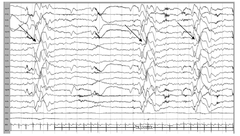
Electroencephalography (EEG) demonstrated multifocal spike and wave paroxysms generalizing during
sleep and bilateral widespread pseudo-periodic slow wave paroxysms with high amplitude (Fig 1).
Atonic-myoclonic seizures were recorded.
Brain magnetic resonance imaging (MRI) revealed no abnormalities but was performed under suboptimal
conditions.
No cells or antimeasles antibodies were detected in cerebrospinal fluid.
Further testing for metabolic disorders was unremarkable.
Diagnosis
Subcortical band heterotopia (double cortex)
Discussion
Subcortical band heterotopia (double cortex) is a neuronal migration disorder in which bands of gray matter
are interposed in the white matter beneath the cortex. The gene for this malformation is on the X chromosome
termed as doublecortin. Individuals with band heterotopia exhibit a broad clinical spectrum, ranging from
asymptomatic to intractable seizures and intellectual impairment. Subcortical band heterotopia (SBH) has a
sex-linked inheritance and more than 90% of the affected individuals are females. Affected individuals
typically present with epilepsy and a variable degrees of mental retardation. Seizures often start in the
first decade of life. They may progress to multiple seizure types and are usually refractory to medication.
Neurological examination may be normal, but dysarthria, hypotonia, poor fine motor control, or, rarely, a
pyramidal syndrome may be present. Interictal EEG findings correlate with seizure types and severity, and
usually include multifocal and generalized epileptiform discharges as well as generalized slowing
Interictal EEG shows frequent and independent multifocal epileptiform discharges (MISWs) over the right
parietal, right temporal, left temporal, bifrontal, and other regions, as well as 500-mcv generalized delta
slowing.
Interictal waking EEG demonstrates a background of generalized theta and delta slowing.
During sleep, there are several second runs of generalized slow spike–wave and continued MISW.
4. A 5-month-old male infant with right-sided facial twitching and generalized tonic-clonic
seizures.
Case presentation
A male infant presented with right-sided facial twitching with secondary right-sided unilateral and
generalized tonic-clonic seizures at 5 months of age.
His parents were unrelated and healthy.
The patient had been born after 39 weeks and 2 days of gestation, and his intrauterine period and birth
were uneventful.
On the basis of his facial appearance, he was suspected to have trisomy 21, and this was later confirmed
by chromosomal G-band staining (47,XY, þ21).
He showed no asphyxia at birth; however, immediately after birth, stridor, hypotonia, and poor sucking
were observed.
Smile and visual pursuit were observed at 2 months of age, but these were lost after the onset of
seizures.
At 5 months of age, the patient still had no head control.
Investigations
All laboratory data, except that for liver enzymes, were within normal ranges.
Failure to thrive, hypotonia, left-sided flaccid paresis, stridor, saddle nose, macroglossia, umbilical hernia, bilateral simian creases, and bilateral fifth fingers shortening and incurved were observed.
Under ultraviolet (UV) light, irregular, streaky areas of hypopigmentation were observed along the lines of Blaschko on the anterior right chest
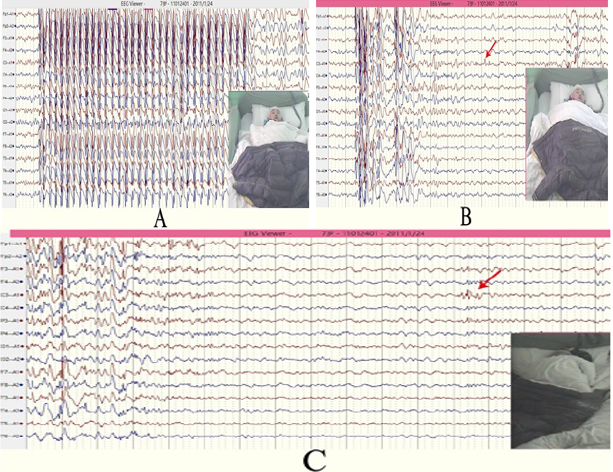
The Interictal electroencephalographic (EEG) findings showed frequent spikes, sharp waves, and spike and wave complexes in the right occipital, parietal, and posttemporal regions (Fig. 1A).
Ictal EEG showed sharp wave bursts in the right-middle temporal region, which developed into polyspikes, and polyspike and wave bursts, involving the right cerebral hemisphere and spreading into both hemispheres (Fig. 1B).
During the clinical course of his epileptic seizures, he showed neither infantile spasms nor hypsarrhythmia.
Brain magnetic resonance imaging (MRI) scans showed focal cortical dysplasia in the right parieto-occipital area, with broad gyri and shallow sulci: axial views show asymmetry due to mild enlargement of the right cerebral hemisphere and ipsilateral enlargement of white matter.
Figure 1. (A) Interictal EEG at age 6 months showing frequent spikes, spike and wave complexes, and high-voltage slow waves in the right occipital and posttemporal regions. (B) Ictal EEG at 6 months of age showing a spike burst in the right-middle temporal region, preceded by rightsided facial twitching, and spreading into the right hemisphere with left unilateral seizures.
Diagnosis
Lateralized periodic discharges in insular status epilepticus
Discussion
In this infant with trisomy 21, hypopigmented patches and depigmentation along the lines of Blaschko were observed. This is a characteristic finding of hypomelanosis of Ito. The patient developed refractory epilepsy due to hemimegalencephaly, predominantly in the right parieto-occipital area. Although some patients with trisomy 21 suffer from epilepsy and febrile seizures, and develop West syndrome during infancy. However, it has been demonstrated that hypomelanosis of Ito can be associated with refractory epilepsy, as well as cortical dysplasia and hemimegalencephaly. Hypomelanosis of Ito is caused by a nonheritable mutation. Hypomelanosis of Ito is often associated with chromosomal abnormalities. Epilepsy in patients with hypomelanosis of Ito is caused by various cortical dysplasia, including hemimegalencephaly. Hemimegalencephaly is most often an isolated congenital abnormality, but it is sporadically associated with neurocutaneous syndromes, such as linear nevus sebaceous syndrome, tuberous sclerosis, hypomelanosis of Ito, and, occasionally, neurofibromatosis.
Interictal electroencephalographic (EEG) findings show frequent spikes, sharp waves, and spike and wave complexes in the right occipital, parietal, and posttemporal regions.
Ictal EEG shows sharp wave bursts in the right-middle temporal region, which developed into polyspikes, and polyspike and wave bursts, involving the right cerebral hemisphere and spreading into both hemispheres.