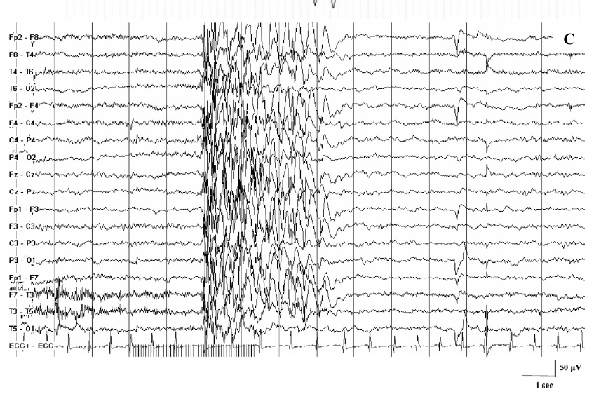
1. Elderly male with repetitive episodes of rising epigastric sensations
Case presentation
A previously healthy 60-year-old man presenting with a 12-month history of repetitive episodes of rising
epigastric sensations was admitted.
He described the episode as a stream of hot gas initially localized to the epigastric region, rising to
the larynx through the thoracic cavity, and ending with a loud gasp.
The entire process lasted for about 1 to several minutes.
The patient experienced this symptom almost every day, and the frequency had increased from once a day
to three to seven times a day.
The symptoms usually occurred during daytime, but sometimes, he was awakened from sleep at night due to
the abnormal sensations in the stomach region.
The patient remained conscious during the episode and had detailed memory of the episode.
The patient had not sought medical attention for the condition until his normal daily activities were
disrupted by the highly frequent occurrence of the episodes.
The patient first visited the gastrointestinal clinic as the symptom was felt in the stomach region.
However, medical treatment for improving the gastrointestinal functions had failed to relieve his
symptoms.
The patient had no history of febrile convulsions, head trauma, pre-existent infection, or
cerebrovascular disease, and also denied having a positive family history.
Investigations
Extensive gastrointestinal investigations, including esophagogastroduodenoscopy, had been carried out,
and no abnormalities were detected.
Investigations such as long-term ECG, heart ultrasonography, coronary artery radiography, and lung
function tests failed to reveal any abnormalities.
Neurological examinations, psychological tests, and other related examinations did not yield any
positive findings. Given the frequency and times of the seizures, epilepsy was suspected.
As routine electroencephalography has a short testing time and a low positive rate, a 2 h video
electroencephalography (V-EEG) was performed, which showed no abnormal activities in the background or
during the interval phase.
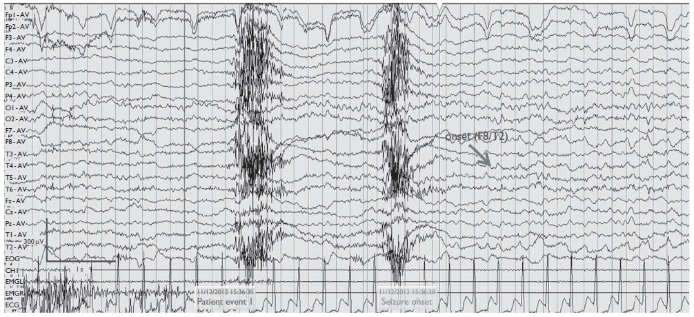
However, during the episode, a slow-wave and sharp-wave focus originated from the right
fronto–centro–temporal region and extended to all leads placed on various parts of the scalp in the
right hemisphere.
MRI scan also revealed sclerosis in the right hippocampus and enlargement of the right temporal horn.
The classical clinical seizure recorded with V-EEG shows the θ-rhythm amplitude in the right
fronto–centro–temporal region, which gradually increases to the high-amplitude δ activity in all leads
of the right hemisphere.
Diagnosis
On the basis of the patient’s medical history and findings of V-EEG and brain MRI, the patient was diagnosed
with Mesial temporal lobe epilepsy (MTLE) associated with simple partial seizures of the autonomic nerves.
Discussion
Abdominal epilepsy is an uncommon syndrome, and the seizure activity results in gastrointestinal symptoms.
MTLE is the most common cause of intractable epilepsy in adults. MTLE is often associated with
viscerosensory and affective manifestations such as a rising epigastric sensation. The ‘early insult’ is
followed by a prolonged latent period during which the patient experiences an abdominal aura, as a sense of
burning with a rising epigastric sensation along with emotional disturbances, such as fear and autonomic
symptoms. MTLE presenting with only visceral manifestations, but without progression to impairment of
consciousness or other manifestations, is extremely rare. This case had several unusual features that are
not commonly observed in cases of abdominal epilepsy. First, the patient presented at the age of 60 years,
which is relatively late for the onset of simple partial seizures with abdominal symptoms in general; this
type of seizure is generally seen in young adults and children. Second, the patient’s symptoms were limited
to epigastric sensations, without vomiting or other autonomic symptoms. In the presence of visceral and
autonomic symptoms without disturbances of consciousness, the patient should undergo thorough evaluation,
including physical and neurological examination and laboratory studies, to exclude other etiologies. V-EEG
should be performed to identify abnormalities specific to epilepsy. If clinical and V-EEG findings are
consistent with abdominal epilepsy, brain MRI should be performed. In this case, the nature of the episode
with the sudden onset, short duration, and occasional occurrence at night helped rule out diseases of the
gastrointestinal tract. Early investigation with V-EEG and brain MRI could help diagnose the condition at an
earlier stage and preclude expensive and invasive interventions.
EEG Tips:
Patients with abdominal epilepsy usually have specific EEG abnormalities, particularly of a temporal
lobe seizure disorder.
During episode of seizure, a slow-wave and sharp-wave focus can originate from fronto–centro–temporal
region and extend to all leads.
2. 13 year old girl presented with pharmacoresistant epilepsy with seizures
Case presentation
A 13-year-old right-handed girl was referred for pharmacoresistant epilepsy with seizures occurring once
or twice per week.
Her seizures had started at 10 years old.
She had sporadic generalized seizures which had increased gradually to 2 per week by 11 years of age.
She had been on antiepileptic medication since 11 years of age.
Generalized seizures stopped with lamotrigine; however, after starting lamotrigine, she began to have
another seizure type, complex partial seizure, composed of abdominal discomfort and mouth automatisms.
Multiple medications failed to control this type of seizure.
There is no family history of seizures.
Investigations
Brain magnetic resonance imaging (MRI) revealed no structural abnormality. Fluorodeoxy glucose positron
emission tomography (FDG-PET) imaging showed mild focal hypometabolism in the left anterior and mesial
temporal lobes.
An ictal–interictal subtraction single photon emission computed tomography (SPECT) study injected 65 s
after seizure onset demonstrated an area of hyperperfusion in the left anterior temporal region during a
seizure with stereotypical focal seizures.
Functional MRI (fMRI) showed left hemispheric language dominance, and neuropsychological evaluation
revealed that her verbal memory had declined.
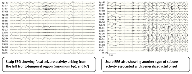
Scalp video-EEG monitoring revealed generalized spike and wave complexes and continuous slow and
interictal spikes in the left frontotemporal region (Fp1 and F7).
After antiepileptic drug (AED) reduction, 21 stereotypical focal seizures were recorded, characterized
by abdominal aura followed by mouth automatism.
In these seizures, EEG seizure activities arose from the left frontotemporal region (maximum Fp1 and
F7).
In addition, 10 nonclinical seizures were recorded associated with generalized ictal onset.
Diagnosis
Based on these evaluations, it was concluded that she had well-controlled generalized epilepsy and
uncontrolled non-lesional left frontal or temporal lobe epilepsy.
Discussion
In the classification of epilepsy, focal epilepsy and generalized epilepsy are two different types, and the
treatment strategies are also different. It has been previously reported that both conditions may coexist in
the same patient and thereby present unique diagnostic and therapeutic challenges. This is a case of a
patient presenting with coexisting left mesial temporal lobe epilepsy and idiopathic generalized epilepsy
(IGE). Intracranial EEG recording demonstrated two types of clearly different seizures: generalized ictal
onset and left mesial temporal onset. In addition, two types of interictal spike distribution, including the
left mesial temporal region and generalized spikes, also supported the coexistence of left mesial temporal
lobe epilepsy and IGE. It has been previously reported that patients with IGE could show EEG focal features.
Furthermore, the coexistence of focal epilepsy and IGE is
rare, and the incidence rate is estimated to be 0.2% of all epilepsy patients and <1% of the IGE population.
This similarity of EEG findings and rarity may cause difficulties in identifying the coexistence of
different seizure patterns. This situation tends to be seen in the pediatric population specifically,
and most focal epilepsy associated with IGE is mesial temporal lobe epilepsy as seen in the present
case. Good seizure control can be achieved with appropriate treatment such as a broad-spectrum
antiepileptic drug or surgical treatment for cases of medically intractable focal seizures coexisting
with IGE. Previous studies suggested that coexistence of generalized epilepsy should not rule out the
consideration of focal epilepsy. It is important to appropriately diagnose coexisting focal and
generalized epilepsy for both medical and surgical management.
EEG Tips:
In cases of coexisting left mesial temporal lobe epilepsy and IGE, intracranial electrodes
showed two characteristic ictal and interictal patterns.
Patient may exhibit typical generalized spike-wave and polyspike-wave discharges and focal
bitemporal sharp-wave discharges in sleep.
3. A 21-year-old man with frequent brief episodes of absent mindedness
Case presentation
A 21-year-old man was presented for frequent brief episodes of absent mindedness throughout the
day and generalized convulsions two or three times a month.
The developmental milestones were normal, and the family history was unremarkable.
The first episode of absentmindedness was noted at the age of 11.
The brief episode, lasting 5–6 seconds, occurred many times daily with subtle bilateral
tremor-like hand movements.
Although no prolonged loss of consciousness occurred, the patient could not recall memories and
details occurring during the episode.
At 13 years of age, a generalized convulsion occurred, which lasted 2–3 minutes with the head
deviating to the left side and subsequent postictal drowsiness.
Following this event, generalized convulsions occurred with a fluctuating pattern of once a day
for several days continuously or two or three times per month; the precipitating factors were
noted to be nervousness, fatigue, hunger, playing on the computer, and taking long baths.
Simultaneously, although brief episodes of absent mindedness occurred many times daily as
before, the patient noted that they were more easily triggered by walking several steps.
At 16 years of age, the patient was brought to a medical center, where EEG (including 3 minutes
of hyperventilation and intermittent photic stimulation) and MRI of the brain revealed no
abnormalities.
Investigations
Neurological examination findings were normal.
During hospitalization, the patient underwent video/EEG monitoring.
Initially, the patient was advised to lie on the bed; however, no event could be identified in
the first 48 hours.
Subsequently, the patient insisted that the event was easily triggered by walking; two brief
episodes of absent mindedness were accordingly noted with clinical confirmation based on EEG
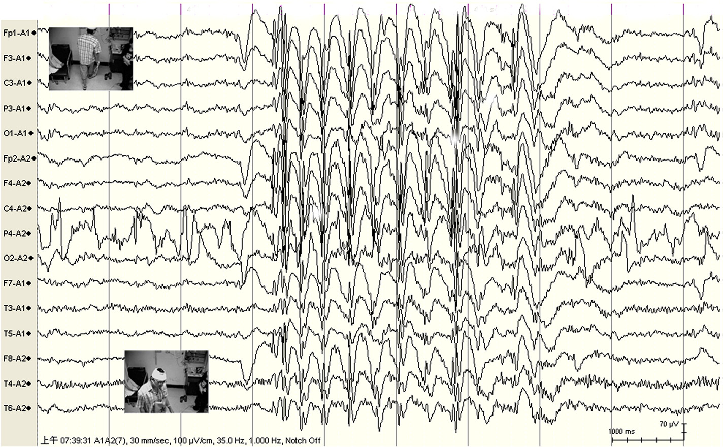
findings of generalized 3-Hz spike-and-wave (SW) complexes lasting 3 and 4 seconds,
respectively, while the patient walked to and fro five to seven steps in the ward.
No postictal confusion could be identified.
The interictal background was normal.
Hyperventilation, intermittent photic stimulation, thinking about walking, imitation of walking
when lying on the bed, and tapping the soles of the feet alternately could not trigger the same
event.
EEG finding of sudden bursts of generalized high-voltage 3-Hz spike-and-wave complexes
for 3–4 seconds.
Diagnosis
Reflex absence epilepsy induced by movement (gait) was diagnosed.
Discussion
The identification of a patient with reflex epilepsy depends on the physician's awareness and the
observations of the patient and witnesses. The epileptogenic trigger must occur often enough in
everyday life so that the patient suspects its relationship to the resulting seizures. This patient
was aware that the absence seizures (ASs) were being more easily triggered by walking several steps.
Video/EEG monitoring verified the trigger mechanism by both clinical and EEG criteria; that is, ASs
consistently followed the action of walking several steps. The recruitment that produces these
seizures need not be confined to physically contiguous brain tissue or fixed neuronal links.
Instead, it may depend on the activity of a function-related network of both established and plastic
links between brain regions that are modified by the effects of factors related to the specific
stimulation. In this patient, the action of walking might have recruited the established links of
the function-related network of ASs. However, the relationship between the plastic links associated
with ASs and walking remain unknown. Although generalized tonic–clonic seizures often develop during
adolescence, the relationship between the simultaneity of generalized convulsions and the trigger
(in this case, gait) of ASs is also unknown.
EEG Tips:
In seizures induced by a specific stimulation, the relatively localized trigger induces
generalized or bilateral EEG abnormalities and seizures.
In gait associated absence seizures there is a sudden burst of generalized high-voltage
spike-and-wave complexes during movement.
4. A 15 year old boy with progressive change in electro-clinical features
Case presentation
A 15 year-old boy with normal development, began to experience episodes characterized by a
sudden vision of “colored spots (red, green, pink) moving in front of me and on the right, like
a flash” at the age of six.
These episodes were all triggered by photic stimuli, such as natural flickering lights, TV or
computer.
EEG studies demonstrated rare interictal spikes or polyspikes with posterior topography, and a
generalized PPR with 3 Hz SW and PSW discharges.
Intermittent photic stimulation (IPS) induced focal seizures which originated from left
temporo-occipital regions and were characterized by visual hallucinations followed by mental
slowing.
Electro-clinical findings suggested an IPOE and he was followed up without antiepileptic drugs
(AEDs).
His mother mother had a single tonic–clonic seizure at age of sixteen and experienced upper limb
myoclonic jerks on awakening.
Investigations
Subsequent video-EEG recordings showed the evolution of electro-clinical patterns over time.
From 9 to 11 years of age, EEG activity was similar to that observed at onset.
More recent EEG recordings (age 11–15) showed progressive change in electro-clinical features:
generalized 3.5 Hz spike-and-wave (SW)/ polyspike-and-wave (PSW) interictal discharges with
progressively more regular and typical morphology replaced the initial focal abnormalities.
An ambulatory 24-h EEG recording even showed activation of these typical generalized 3–3.5
SW/PSW on awakening.
Furthermore, the patient presented a generalized tonic–clonic seizure without any evident
preceding focal sign and also began to experience typical absence seizures.
These absence seizures, recorded during video-EEG monitoring after IPS, consisted of a brief
loss of contact, with staring and perioral myoclonic jerks, and were related to 3.5 Hz
generalized PSW discharge.
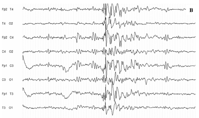
Activation of interictal generalized PSW abnormalities on awakening from an ambulatory
24 h EEG recording at age 15.
Typical photic-induced (18 Hz IPS) absence seizures with staring, brief loss of contact,
and ictal 3 Hz generalized PSW discharge.
Diagnosis
Evolution from idiopathic photosensitive occipital lobe epilepsy to idiopathic generalized epilepsy
(IGE).
Discussion
This case is unique as it demonstrates the natural evolution from exclusively photic-induced
occipital seizures to the progressive emergence of a generalized electro-clinical pattern in an
untreated patient. Although the patient has a tendency towards IGE (supported by the family history)
and that a coincidental association between idiopathic photosensitive occipital lobe epilepsy
(IPOE) and juvenile myoclonic epilepsy (JME) cannot be ruled out, the previous reports of IPOE seen
in families with IGE suggest that these two entities share a common background and support the
hypothesis of their syndromic association. From a pathophysiological standpoint, the thalamocortical
system seems to be involved in the generation of both focal and generalized seizures. In
photosensitive
patients the discharge arises from visual cortex, where magnocellular projections synchronize first
cortical and then subcortical reticular activity. Predominant cortical involvement is reflected by
early visual symptoms, promptly followed by phenomena due to discharge spread, especially to motor
areas. Similarly, early (but likely secondary) involvement of subcortical structures may be
responsible for generalized manifestations such as absences.
EEG Tips:
In patients with syndromic IPOE/ IGE overlap, visual stimuli can trigger abnormal activation of cortical–subcortical network, whose variable involvement can generate different (focal and/or generalized) electro-clinical features in the same family or in the same patient.
A change of electro-clinical features over time could reflect an evolution from a partial to generalized syndrome.