A 22-year-old male was admitted to the hospital due to facial angiofibromas, fibrous cephalic plaque,
ungual fibromas, and asymptomatic renal angiomyolipomas.
Since age six he was suffering from complex partial epilepsy characterized by staring or eye version
(left or right), head and body rotating to the left or right with or without seizures.
Drooling, oro-buccal automatisms, facial expression of fear or anger observed during seizures.
Seizure duration ranged from 3 to 10 minutes; frequency reported was 10~15 episodes per month, often in
clusters of 3~4 successive seizures.
Investigations
Brain magnetic resonance imaging (MRI) and computerized tomography (CT) revealed numerous cortical and
subcortical tubers, white matter radial migration lines and subependymal nodules.
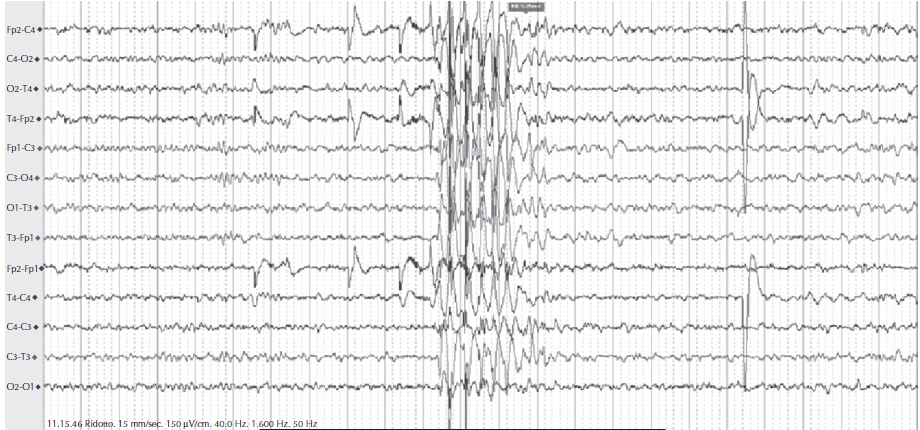
Interictal scalp electroencephalogram (EEG) showed bilateral frontotemporal synchronous slow
spike-and-wave discharges without definite side predominance (Figure 1).
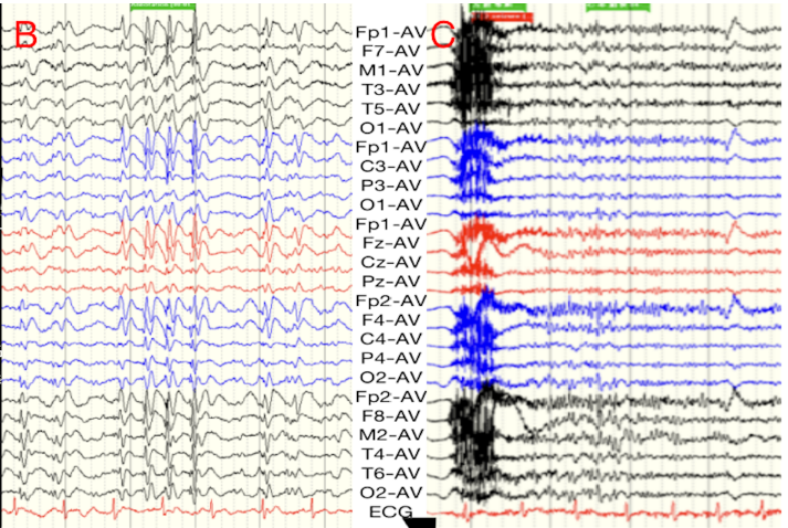
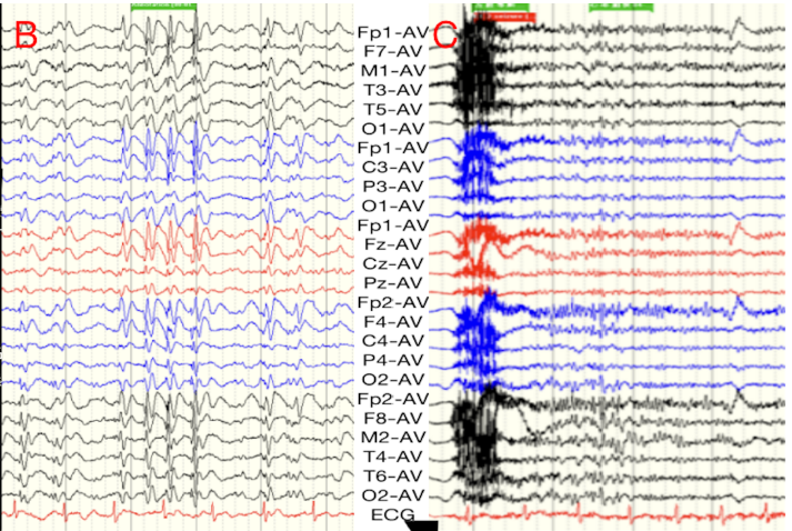
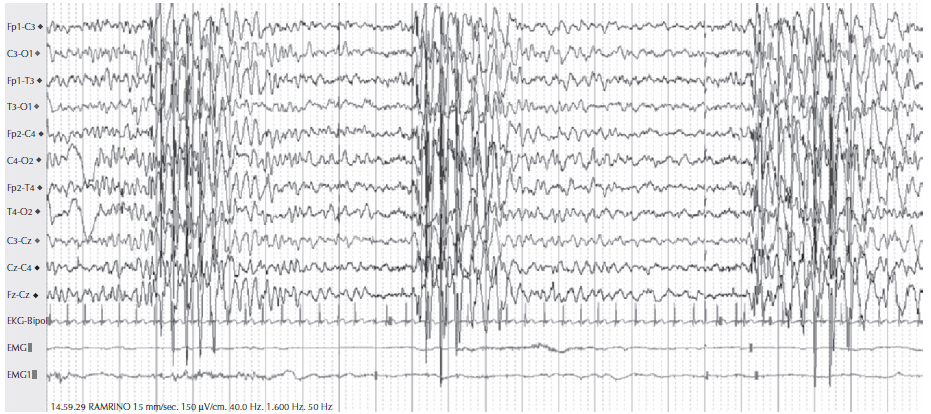
Ictal onsets were poorly localized and appeared generalized and bilaterally synchronous low voltage fast
rhythm (Figure 2).
Valproate (750mg bid), oxcarbazepine (600mg bid) and lamotrigine (50mg qn) were successively added to
patients for antiepileptic treatment, but failed to effectively control seizures.
After pre-surgical evaluation, anterior thalamic nucleus-deep brain stimulation (ANT-DBS) was proposed.
MRI shows two electrodes were implanted into bilateral ANT using standard stereotactic techniques.
The placement of electrodes was determined by Lead-DBS V2 to visually reconstruct the trajectory and
contact position of the electrodes in matrix laboratories (MATLAB).
Regarding seizure severity the LSSS was significantly reduced compared to scores prior of ANT-DBS
(scores of prior vs after ANT-DBS: 63 vs 48).
The QOLIE-31-P total score as well as its subscales improved significantly (total scores of prior vs
after ANT-DBS: 45 vs 50).
Figure 1: Slow spike and wave discharge Figure 2: Low voltage fast rhythm
Diagnosis
The patient was diagnosed with intractable epilepsy caused by tuberous sclerosis (TSC).
Discussion
Seizures were mainly manifested by deep temporal and frontal lobe epilepsy that are suspected to originate in
the limbic system. Neuromodulation is recommended for patients with refractory tuberous sclerosis related
epilepsy (TRE), who are unable to localize epileptogenic nodules after comprehensive preoperative evaluation
or for patients and families who do not agree to resection. Deep brain stimulation of anterior thalamic
nucleus (ANT-DBS) has shown excellent efficacy in the treatment of refractory epilepsy and may be superior
to vagus nerve stimulation (VNS) in epilepsy control and cognitive improvement. ANT-DBS may not achieve the
significant effect of surgical resection, but it is safer and more reversible than surgical resection,
providing a neuromodulation therapy in addition to VNS FOR TRE patients who are unsuitable candidates or
refuse for resection, and more effective than VNS.
EEG Tips:
EEG showed bilateral frontotemporal synchronous slow spike and wave discharge without definite side
predominance
2. Early-Onset Absence Epilepsy Aggravated by Valproic acid
Case presentation
A 3-years-old boy was experiencing stereotyped daily episodes of staring spells associated with brief
isolated jerks of the upper limb.
He was born from non-consanguineous parents after a full term pregnancy.
A simple febrile seizure was reported at the age of 6 months.
He had a family history for febrile seizures (FS) and autosomal dominant IGE, featuring absence seizures
with variable age at onset.
Investigations:
The Interictal EEG recording during wakefulness shows that normal and intermittent photic stimulation
(IPS) was unremarkable.
In a state of drowsiness, a discharge comprising a generalized 3-4Hz spike-wave with interposed fast
polyspikes was observed (Figure 1).
EEG epileptiform discharges were not enhanced in slow sleep.
The video-EEG recording performed in a state of drowsiness showed an absence seizure with a mild clonic
component of the upper limbs and brief upward rotation of the eyes.
VPA treatment was started at a dose of 20mg/kg weight (65.5µg/ml) but brief absences seizure occurred
two to three times daily, therefore VPA dose was increased to 30 mg/kg body weight.
During slow sleep, with VPA treatment, frequent generalized 3-4-Hz spike-and-wave complexes with
interposed fast polyspikes were also observed (Figure 2).
VPA caused an increase in seizures, thus discontinued and LEV at 20mg/kg body weight was added. The boy
did not experience any seizures.
SLC2A1 gene testing was negative.
Figure 1: Generalized 3-4-Hz spike-waves with interposed fast polyspikes coincide with a short
absence seizure with brief upward rotation of the eyes and a mild clonic component of the upper limbs.
Figure 2: During slow sleep, with VPA treatment, the patient showed frequent generalized 3-4-Hz
spike-and-wave complexes with interposed fast polyspikes.
Diagnosis
The patient was diagnosed with early-onset absence seizures aggravated by VPA treatment.
Discussion
Early-onset absence epilepsy (EOAE) includes a heterogeneous group of epilepsies with different
electroclinical features and prognosis. Onset of absence seizures in the first year of life is very rare.
The presence of short absence seizures with early onset and myoclonic seizures, are associated with Glut-1
deficiency syndrome. Absence seizures may start in the first years of life, as the main type of seizure, in
well-defined epileptic syndromes, such as MEI, shown in the video-EEG recording. VPA interfere with brain
glucose metabolism and, thus, exacerbate Glut-1 deficiency, leading to seizure aggravation. The increase of
the seizures may be due to a paradoxical mechanism. The mechanism of seizure worsening did not involve VPA
toxicity, encephalopathy, Glut-1 deficiency or overdosage, and the reason for absence seizure aggravation
remained unclear. Complete control of absence seizures achieved with levetiracetam treatment and the course
was benign, both in terms of seizure control and neuropsychological aspects.
EEG Tips:
Video-EEG in a state of drowsiness helps in the diagnosis of short absence seizure
3. Narcolepsy Mimicking Epileptic Seizures
Case presentation:
An 11-year-old girl presented to hospital because of suspected narcolepsy with unique cataplexy or
incidental epileptic seizures.
She had no medical history or family history of epilepsy.
At age 10, she was started to have recurrent myoclonic attack, manifesting as dropping things from her
hands immediately after hearty laughs, also experienced frequent naps, hypernagogic hallucinations, and
sleep paralysis.
Investigations
• The long-term video-The long-term video-polygraph showed an unprovoked myoclonic attack during lunch,
that was characterized by sudden, brief myoclonic movement mainly involving upper limbs during which she
almost dropped chopsticks from her hand.
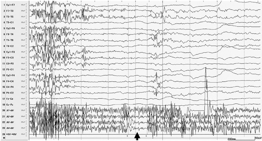
• EMG showed sudden, brief interruption of the sternocleidomastoid, latissimus, and deltoid EMG
discharges lasting for 140 ms, without corresponding EEG changes (Figure 1).
• Narcolepsy was confirmed by positive human leukocyte antigen HLA-DQBI * 0602 and low orexin levels in
CSF
Figure 1: The sternocleidomastoid, latissimus, and deltoid EMG shows sudden, brief interruption
of ongoing muscle activity lasting for 140 ms (arrow); however, the corresponding EEG does not show any
epileptic changes.
Diagnosis
The patient was diagnosed with narcolepsy due to low orexin levels in cerebrospinal fluid (CSF)
Discussion
Narcolepsy is a brain disorder characterized by excessive sleepiness, hypnagogic hallucinations, and sleeps
paralysis, and can occur with or without cataplexy. The association of human leukocyte antigen (HLA) genes,
especially DQB1 * 0602, in patients with narcolepsy with cataplexy suggests that it is an autoimmune
disorder. Childhood epilepsy sometimes present with unique epileptic seizure type that resembles cataplexy.
In the present report, case was described as fine segmental cataplexy resembling myoclonic seizures that
caused the very short-lasting, mild initial phase that did not evolve to the falling phase as well as the
anatomic phase. A subtle inhibitory muscular event could generate a myoclonic-like cataplexy.
Myoclonic-atonic seizures in epilepsy with myoclonic-atonic seizures, massive or subtle myoclonic seizures
in myoclonic epilepsy in infancy or juvenile myoclonic epilepsy, all exhibit similar characteristics to
cataplexy. Children with narcolepsy may have attacks for cataplexy that resembles clonic or myoclonic
seizures.
EEG Tips:
Long-term video-electroencephalography (EEG) helps to differentiate cataplexy from epileptic seizures.
4. Epilepsy Due To Posttraumatic Stress Disorder and Depression
Case presentation:
A 48-year-old male, with right mesial temporal sclerosis was admitted to epilepsy monitoring unit (EMU).
Left side weakness secondary to presumed encephalitis was observed at age 1, but resolved by age 4.
At the age 42, the patient started to have spells, described as asymmetric shaking starting with one leg
and slowly progressing over 2-3 min to involve all four limbs lasting 5-10 min, occurs almost every day.
Approx one year later (at age 43) the patient began to experience diurnal seizures and nocturnal
convulsive seizures, accompanied by tongue biting or urine incontinence and diagnosed by a psychiatrist
with depression and posttraumatic stress disorder (PTSD).
Brain MRI showed evidence of right hippocampal sclerosis.
Despite treatment with combinations of phenytoin, levitiractam, carbamezipine, clobazam lamotrigine,
valproic acid and lacosamide, he continued to have focal epileptic seizures with impaired awareness
evolving to bilateral tonic–clonic seizures at a frequency of 1 every 1–2 months and nonepileptic events
at a frequency of 1–5 per month.
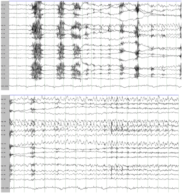
Nine right temporal seizures were recorded during presurgical evaluation, characterized by sudden onset
of behavioral arrest followed by chewing movements lasting for 1 minute and then on two occasions
evolving to bilateral tonic–clonic seizures (Figure 2).
Pathological analysis of the resected tissue revealed dual pathology including hippocampal sclerosis and
focal cortical dysplasia (FCD) type 3A.
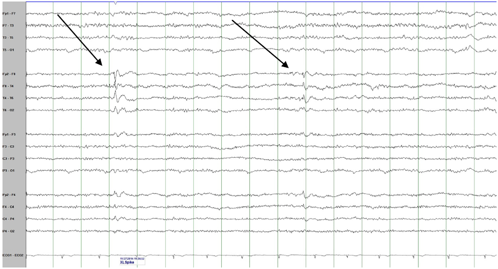
Figure 1: An Interictal EEG showing two examples of Interictal right anterior temporal spikes
Figure 2: Two consecutive epochs of EEG showing the onset and the evolution of the right
temporal ictal rhythm at the onset of one of the patient's recorded epileptic seizures.
Diagnosis
The patient was diagnosed with both drug resistant epilepsy and psychogenic nonepileptic seizures.
Discussion
Drug resistant epilepsy carries increased risk of injury and death. Patients with drug resistant epilepsy
were investigated for surgical options, seeking to achieve seizure freedom. Both conditions coexist with
multiple reports estimating the presence of epilepsy in patients with psychogenic nonepileptic seizures
(PNES) to be less than 10%. Risk factors for developing depression in patients with epilepsy included poor
medication compliance, worse seizure frequency, unemployment and a worse overall quality of life. Depression
can exacerbate seizures and treating it may decrease seizure frequency. The prevalence of depression in
patients known to have PNES (21–60%) is significantly higher than the prevalence rates not only in the
general population, but also in patients with epilepsy whose seizures are controlled, and in patients with
treatment-resistant epilepsy.
EEG Tips:
Video-EEG helps to identify epileptic seizures in patients diagnosed with psychogenic nonepileptic
seizures (PNES).