Tips and Tricks in EEG Diagnosis
1. Biallelic mutations in SZT2 associated with epilepsy and developmental delay
Case presentation
Investigations
Diagnosis
Thick and short corpus callosum and mutations in SZT2 indicating epilepsy and developmental delay.
Discussion
This present case revealed that deficiency results in constitutive mTORC1 signaling. A lack of macrocephaly in patient may be explained by secondary neuronal loss due to recurrent seizures. Macrocephaly associated with non-intractable epilepsy may be a hallmark of SZT2-related epilepsy. Although clinical features of SZT2-related disorders such as various degrees of epilepsy, developmental delay, and macrocephaly are non-specific, the dysmorphic corpus callosum observed in this case with epilepsy. The thick and short shape of the corpus callosum, together with epilepsy, developmental delay and macrocephaly, may be a diagnostic feature of a discernible disorder that results from mutations in SZT2.
EEG TIP
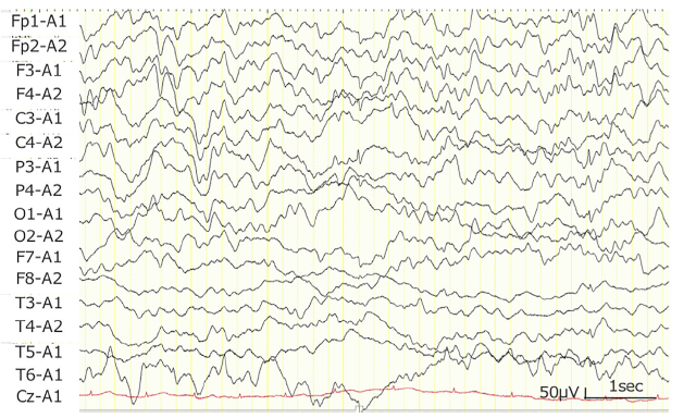
EEG recording helped in focal epileptic discharges which rarely occurred predominantly in the left parietal region.
Nakamura Y, Togawa Y, Okuno Y,et al.Biallelic mutations in SZT2 cause a discernible clinical entity with epilepsy, developmental delay,macrocephaly and a dysmorphic corpus callosum. Brain Dev. 2018 Feb;40(2):134-139.
2. Granular Osmiophilic Elements and mutation in presenilin1 in Early-Onset Alzheimer’s Disease
Case presentation
Investigations
Diagnosis
Diagnosed as early-onset Alzheimer’s disease (EOAD), characterized by rapid cognitive decline associated with myoclonus and seizures.
Discussion
The present case report describes a patient suffering from an early onset of dementia, and bearing a mutation in PSEN 1. The pattern of behavioral disturbances, the adverse response to cholinesterase inhibitors and the benefit from the administration of antiepileptic agents that abolished myoclonic jerks led us to speculate that a similar phenotype and out-come might be attributable to the position of the 2 pathogenic variants in a similar domain of presenilin 1, the gene product. Epilepsy is known to be part of autosomal dominant Alzheimer’s disease phenotype. The cholinergic loss is an early marker of memory deficits in Alzheimer’s disease, and PS1 mutant knock- show impairment of cholinergic modulation in hippocampal plasticity, providing evidence of cholinesterase inhibitor therapy in dementia.
The present case and the patient reported earlier did not harbor variants in CLN1 , it is tempting to speculate that the 2 diseases shared similar pathomechanisms eventually related to a dysfunction of the lysosomal compartment.
EEG TIP
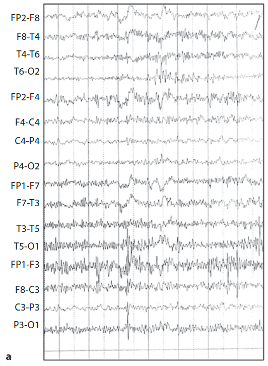
An EEG showed low amplitude pseudoperiodic sequences with spikes and slow waves in the occipital derivations.
Alberici A, Borroni B, Bonato C,et al.. EEG and granular osmiophilic elements in early-onset Alzheimer's disease. Neurodegener Dis. 2011;8(4):259-61.
3. Focal epilepsy with 5,10-methylenetetrahydrofolate reductase (MTHFR) deficiency
Case presentation
Investigations
Diagnosis
Diagnosed as MTHFR deficiency in the presence of epileptic manifestations, including progressive myoclonic or focal epilepsy.
Discussion
Plasma homocysteine is a key biomarker of the disease. Plasma methionine (low to normal) and urine methylmalonic acid (normal) can then further point to the diagnosis of MTHFR deficiency, which can then be confirmed by molecular and/or enzymatic testing.
The present case report underscores the importance of diagnosing MTHFR deficiency in young adults in the presence of epileptic manifestations, including progressive myoclonic or focal epilepsy, especially when associated with central (spasticity, dysexecutive syndrome) and/or peripheral neurological signs (polyneuropathy). The correct diagnosis can lead to specific therapeutic management and the prevention of complications related to hyperhomocysteinemia, and to family screening.
EEG TIP
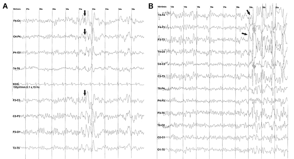
EEG helps in displaying anterior bilateral sharp theta elements and rare sharp slow waves during wakefulness.
Mezouar N, Mochel F, An-Gourfinkel I,et al. Focal epilepsy as the revealing symptom of 5,10-methylenetetrahydrofolate reductase deficiency in a young adult. Rev Neurol (Paris). 2018 Mar;174(3):173-175.
4. Peri-oral myoclonia with absences with multiple facial and upper body myoclonia: Overlap epilepsy syndrome
Case presentation
Investigations
Diagnosis
Peri-oral myoclonia with absence epilepsy was made (POMA) in view of pre dominant myoclonia in the peri-oral region.
Discussion
Perioral myoclonia with absences (POMA) was first described in 1994 by Panayiotopoulos. It is characterized by onset between 2 and 13 years of age, absences with myoclonia of the peri-oral muscles, co-existing generalized tonic-clonic seizures, propensity to absence status and poor response to anti-epileptic drugs. Asymmetric facial twitching and focal EEG abnormalities led to a misdiagnosis of a focal epilepsy in this case. The exact epilepsy syndrome classification may be difficult in this case. Given the predominant peri-oral myoclonia, frequent generalized tonic-clonic seizures, poor response to anti-epileptic drugs, eye closure sensitivity and presence of other myoclonia, a diagnosis of POMA overlapping with EMA was considered.
EEG TIP
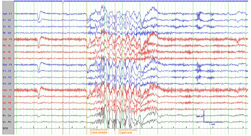
EEG revealed s high amplitude generalized 3.5 Hz spike wave discharges lasting for 3.5 s. which was associated with brief absence with peri-oral myoclonia.
Gulati P, Jain P, Qashqari H,et al. Peri-oral myoclonia with absences with multiple facial and upper body myoclonia: Overlap epilepsy syndrome. J Clin Neurosci. 2018 Aug;54:135-137.